Effect of Sevoflurane on Cognitive Dysfunction
Sevoflurane is a common inhalational anesthetic that plays a critical role in modern surgical procedures due to its favorable properties, including rapid onset and minimal side effects. However, emerging evidence suggests that exposure to sevoflurane may have negative effects on cognitive function. In experimental animal models, sevoflurane has been linked to cognitive dysfunction1, which may manifest as post-operative cognitive dysfunction (POCD) in elderly human patients and neurobehavioral abnormalities in younger subjects. This cognitive impairment, which can persist for weeks or even months post-surgery, raises significant concerns, particularly in aging populations who are already at higher risk for neurodegenerative conditions such as Alzheimer’s disease. Sevoflurane has also been shown to induce neuronal apoptosis and decrease adaptability in neonatal rats.2 Understanding the mechanisms through which sevoflurane induces cognitive dysfunction enables the development of strategies to minimize its impact and improve patient outcomes in surgical settings.
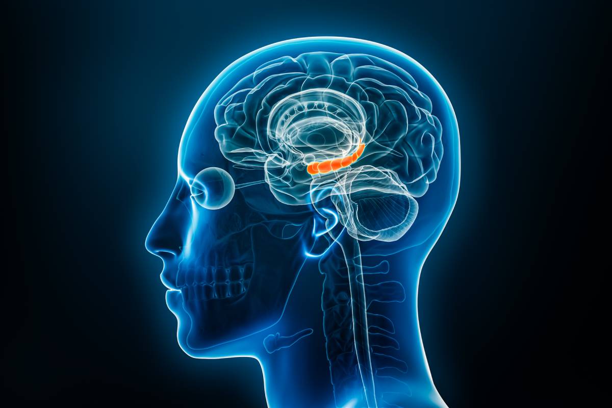
The hippocampus is a critical neuronal structure well known to be involved in learning and memory.3 It is also an important target of general anesthetics. In 2016, researchers performed in vivo experiments on neonatal rats (n=88) to study the effect of sevoflurane anesthesia on hippocampal synaptic plasticity and, subsequently, learning and memory. 30 rat pups received 3% sevoflurane treatment for 1 hour, while another 28 received treatment for 6 hours. The remaining 30 pups served as the control group. Synaptic vesicle-associated proteins and dendrite spine density were assessed using Golgi staining, transmission electron microscopy (TEM), and western blotting. The researchers found neonatal exposure to sevoflurane treatment for 6 hours resulted in reduced spine density of apical dendrites and elevated expression of synaptic vesicle-associated proteins (SNAP-25 and syntaxin). Rats exposed to sevoflurane for 6 hours also performed worse than their counterparts in the Morris water maze and novel-object recognition tests, behavioral tasks meant to measure learning and memory performance. In the group who had received sevoflurane for 1 hour, significantly less structural and functional damage was observed in the hippocampus. The results demonstrate sevoflurane anesthesia may time-dependently induce cognitive dysfunction by deteriorating hippocampal function.4
A similar study on sevoflurane and its effects on hippocampal neurons was conducted on 42 adult rats. Results of an open field test showed decreased locomotor performance for both male and female rats treated with 3% sevoflurane, compared to their control counterparts. However, there was only a significant difference between the experimental and control groups at one day post-anesthesia, but not at 30, 60, or 90 days, indicating sevoflurane inhibits short-term locomotor activities but has little effect on long-term movement.5 For their molecular analysis, the researchers collected data on the cAMP response element binding (CREB) protein, a key gene that mediates downstream transcription imitation factors and regulates neuronal survival. Sevoflurane is known to exert its anesthetic effects through inhibition of hippocampal NMDA receptors, which are heavily connected to CREB protein signaling pathways. Western blotting revealed phosphorylation of CREB was significantly decreased at one week following treatment, whereas no distinct difference was detected in the rat hippocampus at three months post-anesthesia.5 Sevoflurane was also shown to increase levels of Caspase-3 and Caspase-8, death proteases that are crucial mediators of apoptosis.6 Collectively, the results suggest that sevoflurane may induce cognitive dysfunction through inhibiting CREB signaling pathways, which in turn blocks NMDA receptor function.
In general, the existing literature suggests that high levels of exposure to sevoflurane anesthesia may pose risks to cognitive function, possibly through its impact on the hippocampus. Murine studies indicate exposure to sevoflurane can cause neuronal apoptosis and structural damage to the hippocampus. These changes may contribute to post-operative cognitive dysfunction and other neurological deficits concerning learning and memory. However, it is important to note that many of these effects may be short-term, with many individuals recovering their lost cognitive function over time. Nonetheless, the possible time-dependent nature of these effects does not take away from the need for continued research to better understand the duration and reversibility of sevoflurane’s effects on cognitive dysfunction.
References
- Bekker, Alex Y., and Edwin J. Weeks. “Cognitive Function after Anaesthesia in the Elderly.” Best Practice & Research Clinical Anaesthesiology, vol. 17, no. 2, June 2003, pp. 259–72. https://doi.org/10.1016/S1521-6896(03)00005-3
- Zheng, S. Q., et al. “Sevoflurane Causes Neuronal Apoptosis and Adaptability Changes of Neonatal Rats.” Acta Anaesthesiologica Scandinavica, vol. 57, no. 9, Oct. 2013, pp. 1167–74. https://doi.org/10.1111/aas.12163
- Howland, John G., and Yu Tian Wang. “Chapter 8 Synaptic Plasticity in Learning and Memory: Stress Effects in the Hippocampus.” Progress in Brain Research, edited by Wayne S. Sossin et al., vol. 169, Elsevier, 2008, pp. 145–58. https://doi.org/10.1016/S0079-6123(07)00008-8
- Xiao, Hongyan, et al. “Learning, Memory and Synaptic Plasticity in Hippocampus in Rats Exposed to Sevoflurane.” International Journal of Developmental Neuroscience, vol. 48, Feb. 2016, pp. 38–49. https://doi.org/10.1016/j.ijdevneu.2015.11.001
- Xie, H., et al. “The Gender Difference in Effect of Sevoflurane Exposure on Cognitive Function and Hippocampus Neuronal Apoptosis in Rats.” European Review for Medical and Pharmacological Sciences, vol. 19, no. 4, Feb. 2015, pp. 647–57.
- Porter, Alan G., and Reiner U. Jänicke. “Emerging Roles of Caspase-3 in Apoptosis.” Cell Death & Differentiation, vol. 6, no. 2, Feb. 1999, pp. 99–104. https://doi.org/10.1038/sj.cdd.4400476