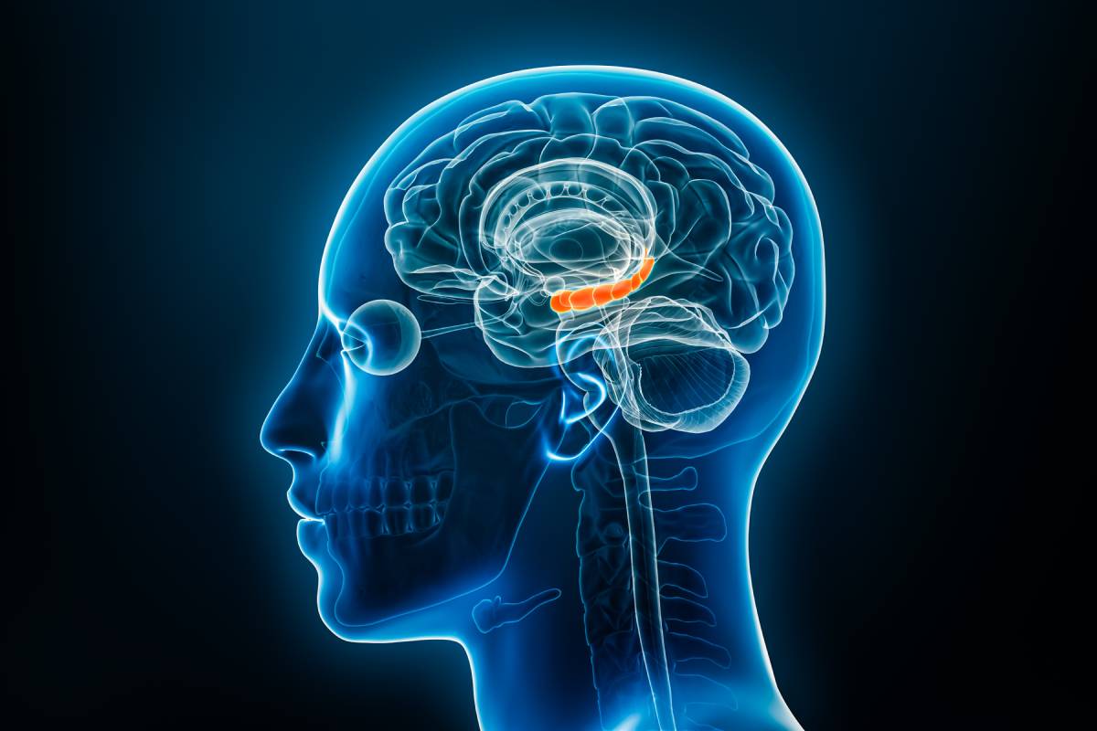
A genicular nerve block is a minimally invasive procedure used to diagnose or treat chronic knee pain, particularly in cases where other treatments such as medications or physical therapy have not provided adequate relief. This technique targets the genicular nerves—small sensory nerves that transmit pain signals from the knee joint to the brain. By blocking these nerves, the procedure can reduce or eliminate pain, allowing for improved mobility and quality of life 1.
The genicular nerve block is most commonly used for patients suffering from chronic osteoarthritis of the knee, especially those who are not candidates for knee replacement surgery. It is also used for individuals experiencing persistent post-surgical knee pain or pain following knee trauma. In some cases, the block is used as a diagnostic tool to determine whether a patient is likely to respond well to a longer-term treatment such as genicular nerve ablation 2,3.
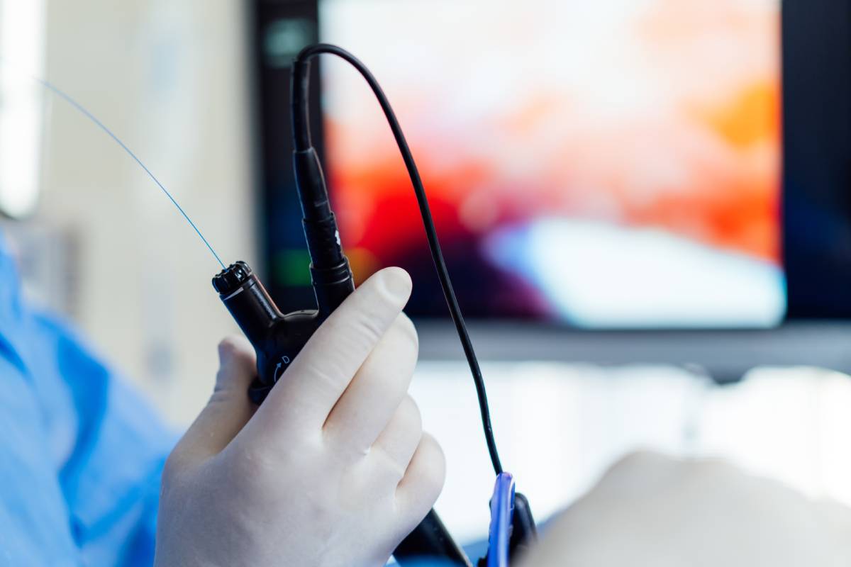
During the procedure, a healthcare provider injects a local anesthetic, often combined with a steroid, near the genicular nerves around the knee. Using fluoroscopy or ultrasound, the provider ensures that the needle is accurately placed; the injected anesthetic temporarily blocks the transmission of pain signals from the knee to the brain.
Genicular nerve blocks are typically performed by pain management specialists, anesthesiologists, or other physicians with specialized training. These providers have expertise in musculoskeletal and nervous system anatomy and the use of image-guided procedures for injections near nerves to ensure precision and safety. The procedure is typically carried out on an outpatient basis and takes less than half an hour. Most patients report only mild discomfort during the injections and can return home shortly afterward. Driving is usually not recommended immediately after the procedure, so arranging for transportation is advised 1,4–6.
Many individuals experience significant pain relief within hours of the procedure. The duration of relief varies depending on the individual, but it typically lasts several days to a few weeks for diagnostic blocks. If the patient experiences meaningful relief, this can indicate that a more permanent treatment, such as radiofrequency ablation of the genicular nerves, may be effective. In such cases, the nerves are targeted with heat energy to disrupt pain signals more permanently 4,7,8.
The genicular nerve block is considered safe, but like all medical procedures, it carries some risks. These include temporary soreness at the injection site, bleeding, infection, or allergic reaction to the medications used. Rarely, the procedure may not provide any pain relief, or the relief may be short-lived. Proper patient selection and imaging guidance significantly reduce the risk of complications 1,9.
The genicular nerve block is an effective, low-risk option for diagnosing and managing chronic knee pain, especially for those seeking alternatives to surgery. When successful, it can lead to longer-term solutions and help restore a patient’s mobility, comfort, and quality of life.
References
- Genicular Nerve Block. Cleveland Clinic https://my.clevelandclinic.org/health/treatments/24823-genicular-nerve-block.
- OSC, O. Diagnostic Genicular Nerve Blocks for Knee Pain. Orthopaedic and Spine Center of Newport News https://www.osc-ortho.com/blog/diagnostic-genicular-nerve-blocks-for-knee-pain/ (2021).
- Genicular Nerve Block | Pain Specialists of America. https://www.psadocs.com/services/genicular-nerve-block/.
- Hasoon, J., Orhurhu, V. J. & Yazdi, C. Genicular Nerve Blocks for the Management of Chronic Knee Pain Related to Osteoarthritis – A Case Series. Orthop Rev (Pavia) 16, 126046. DOI: 10.52965/001c.126046
- NYSORA. Genicular Nerve Blocks. NYSORA https://www.nysora.com/techniques/lower-extremity/nysora-com-genicular-nerve-blocks/ (2021).
- Genicular Nerve Blocks – AOA Orthopedic Specialists. https://www.arlingtonortho.com/conditions/knee/genicular-nerve-blocks/ (2018).
- Li, W., Xu, F., Chen, F., Cao, L. & Bao, X. Effect of Genicular Nerve Block (GNB) on Pain in Lesions of the Knee Joint: A Meta-Analysis of Randomized Controlled Trials. JPR 18, 511–522 (2025). DOI: 10.2147/JPR.S503937
- Konya*, Z. Y., Akin Takmaz, S., Basar, H., Baltaci, B. & Babaoglu, G. Results of genicular nerve ablation by radiofrequency in osteoarthritis-related chronic refractory knee pain. Turk J Med Sci 50, 86–95 (2020). DOI: 10.3906/sag-1906-91
- Ferguson MD, K. & Kalia MD, H., MPH, FIPP, FACPM. Complications of Genicular Nerve Blocks and Ablations. in Complications of Pain-Relieving Procedures 387–391 (John Wiley & Sons, Ltd, 2022). DOI: 10.1002/9781119757306.ch45.