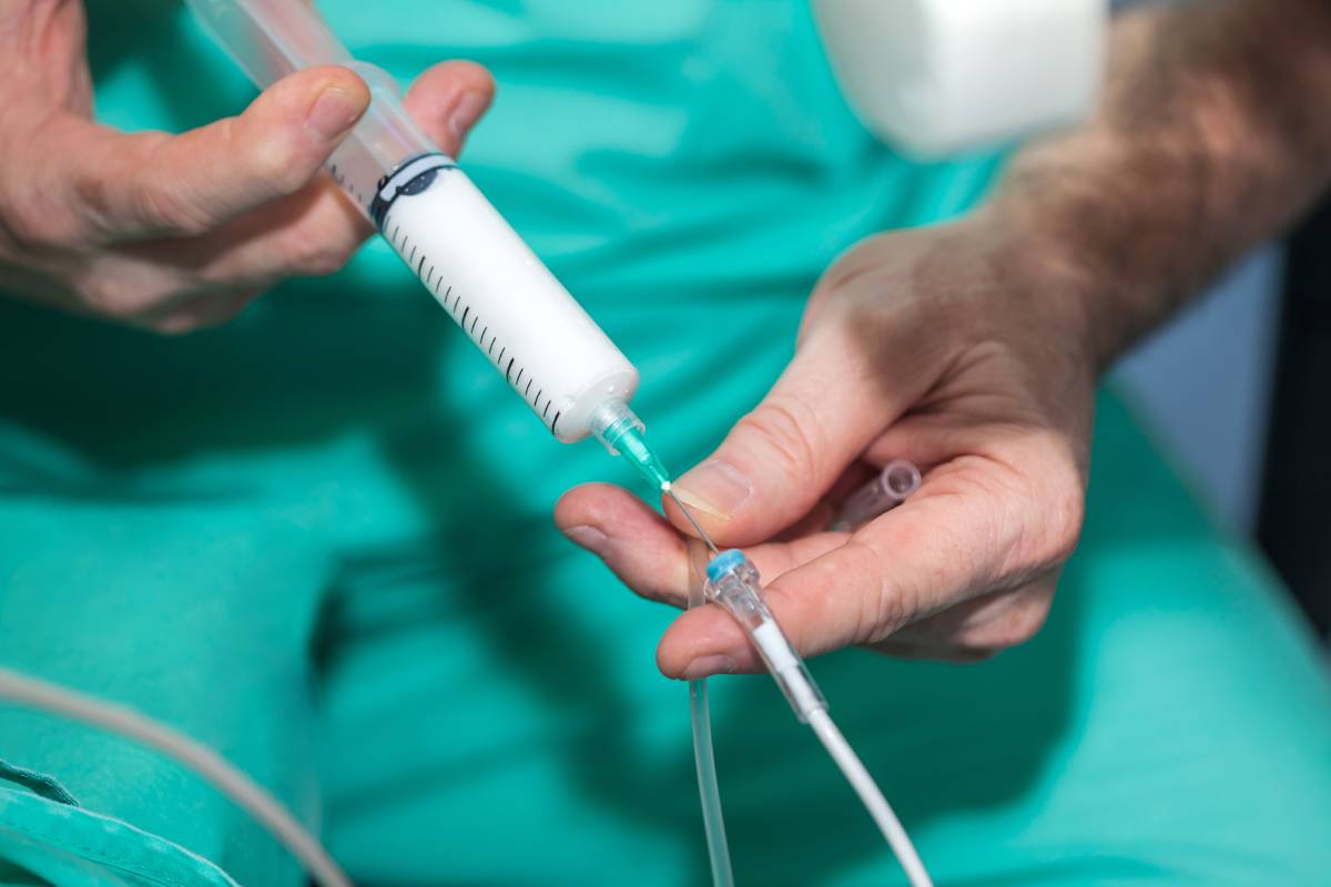
Precautions Addressing Substance Use Disorder in Anesthesiologists

According to a study conducted in 2020, at least 1.6% of anesthesiologists are likely to suffer from substance use disorder during their careers [1]. Alcohol, opioids, and anesthetics are some of the substances that these physicians abuse most often [1]. Were substance use disorder considered an occupational hazard, anesthesiologists would rank among the most endangered workers in the United States [1]. 

Notwithstanding an individual’s profession, substance use disorder exposes its sufferers to severe risks. It is particularly dangerous for anesthesiologists, given the responsibility that they wield over the lives and health of other people [2]. Substance use disorder can impede an anesthesiologist’s training and result in severe professional consequences, such as failure to become certified in a subspecialty or to complete residency [2]. With the trajectory of anesthesiologists’ own lives – as well as the lives of others – in danger, the importance of curbing substance use disorder among them cannot be understated.  

Different types of policies serve to address this problem, with varying degrees of success. For instance, educational programs have been created to educate anesthesiologists about how to identify the disorder and intervene when they recognize it in their peers [2]. Identification can be difficult, given how anesthesiologists are cognizant of the signs of substance use disorder and thus more aware of how to hide them from others [3]. A pivotal warning sign to look out for is a change in the suspected individual’s functional capacity: substance abuse impedes people’s ability to carry out certain duties [3].  

Other warning signs are the replacement of a syringe or ampule’s contents with saline and the taking of narcotics from disposal containers [3]. Anesthesiologists with substance use disorder may also report that a case is opioid-based but then only administer beta-blockers and inhalational agents to the patient, taking the opioid medication for their own use [3]. To prevent these actions, some medical facilities conduct regular inspections of dispenser transactions and anesthetic records [2]. Meanwhile, to combat the removal of substances from waste, greater security measures may be wise. 

A more recent innovation has been the randomized substance testing of anesthesiologists, among other personnel, in medical institutions [2]. This strategy is not widely used – it appears to occur in a limited amount of civilian hospitals – but a study of anesthesiology residents at Massachusetts General Hospital suggests that it could be helpful in disincentivizing substance abuse and identifying it when it does occur [2, 4]. The experiment reported no cases of substance use over its 1,002-resident-year testing period, compared with four cases during the previous 719 resident years [4]. This was a limited study, however, so further research must occur to fully understand the beneficial effects of drug testing, if any, on substance use disorder among anesthesiologists and other physicians [4]. 

While submitting anesthesiologists to randomized testing may help identify those who suffer from substance abuse, the pathways to voluntary confession should also be improved to give physicians who wish to seek help an easy way to do so. It typically takes years for substance abuse to become apparent [5]. Giving anesthesiologists an option to disclose their disorder to a helpful, responsible authority could help challenge the stigma associated with the disorder and, ultimately, encourage anesthesiologists to openly seek help [6].  

Because of the incompleteness of current strategies, such as educational and surveillance programs, a multi-faceted approach that employs several of the aforementioned precautions may be the best way to address substance use disorder among anesthesiologists [2]. Through trial and error, medical institutions may be able to combat this problem and improve the health of their patients and their personnel. 

References 

[1] D. O. Warner et al., “Substance Use Disorder in Physicians after Completion of Training in Anesthesiology in the United States from 1977 to 2013,” Anesthesiology, vol. 133, p. 342-349, August 2020. [Online]. Available: https://doi.org/10.1097/ALN.0000000000003310

[2] E. O. Bryson, “The opioid epidemic and the current prevalence of substance use disorder in anesthesiologists,” Current Opinion in Anesthesiology, vol. 31, no. 3, p. 388-392, June 2018. [Online]. Available: https://doi.org/10.1097/ACO.0000000000000589

[3] L. G. Lefebvre and I. M. Kaufmann, “The identification and management of substance use disorders in anesthesiologists,” Canadian Journal of Anesthesia, vol. 64, p. 211-218, November 2016. [Online]. Available: https://doi.org/10.1007/s12630-016-0775-y

[4] M. G. Fitzsimons et al., “Reducing the Incidence of Substance Use Disorders in Anesthesiology Residents: 13 Years of Comprehensive Urine Drug Screening,” Anesthesiology, vol. 129, p. 821-828, October 2018. [Online]. Available: https://doi.org/10.1097/ALN.0000000000002348

[5] D. Volquind et al., “Occupational Hazards and Diseases Related to the Practice of Anesthesiology,” Brazilian Journal of Anesthesiology, vol. 63, no. 2, p. 227-232, March-April 2013. [Online]. Available: https://doi.org/10.1016/j.bjane.2012.06.006

[6] S. J. S. Bajwa and J. Kaur, “Risk and safety concerns in anesthesiology practice: The present perspective,” Anesthesia Essays and Researchers, vol. 6, no. 2, p. 227-232, March-April 2013. [Online]. Available: https://doi.org/10.1016/j.bjane.2012.06.006.

Safety and Quality Ratings for ASCs 

Ambulatory surgery centers (ASCs), modern healthcare facilities focused on providing same-day surgical care (including preventive and diagnostic procedures), have emerged as key players in ensuring safe, high quality, and cost-effective health care delivery 1. Indeed, with a strong record of positive patient outcomes, they have reshaped the outpatient experience for millions of American patients by providing them with a convenient alternative to hospital-based procedures 2. However, their safety and quality is important to monitor in order to ensure that patient outcomes are not negatively impacted. Several organizations, both public and private, provide safety and quality ratings for ASCs to this end.

Capitalizing on recent advances in surgical and pain management techniques, ASCs today regularly and safely perform most outpatient surgery procedures. In addition, since ASCs often specialize in certain specialties and/or procedures, they are able to place additional focus on patient experience and safety. In so doing, similar to hospital operating rooms, surgeons, nurses and medical professionals in ASCs adhere to a strict set of protocols  3

The safety and quality of ASCs is ensured by close monitoring. Like hospitals, ASCs must abide by a number of laws and regulations. Most ASCs, for example, are Medicare-certified, and a large number are accredited by independent agencies that provide safety and quality ratings after a thorough screening and inspection process. 

In order to maintain a safe and sanitary environment, every ASC needs to establish and maintain procedures for preventing infections. The Centers for Disease Control (CDC) recommend contracting teams specializing in infection prevention 4 which can manage an ASC’s infection prevention program in a facility-tailored way. Meanwhile, individual ASCs may also utilize standardized surveys established to rate their own safety and implement changes accordingly 5. The national Agency for Healthcare Research and Quality (AHRQ), to this end, created the Ambulatory Surgery Center Survey on Patient Safety Culture. The survey is specifically designed for ASC staff, focusing on their own assessments of patient safety in their facility. 

Similarly, ASCs must conduct regular, comprehensive ratings of the safety and quality of care they provide to their patients. Today, both Medicare’s Ambulatory Surgical Center Quality Reporting (ASCQR) program and a program coordinated by the ASC Quality Collaboration (ASC QC) serve as robust, open-access ASC quality reporting programs 6

The ASC QC was created in 2006 as a cooperative effort between a number of different organizations in order to begin to develop standardized ASC quality measures 8, concentrating on both outcome and process measures. To date, nearly 2,000 ASCs have voluntarily participated in the reporting of its 11 distinct ASC performance measures.  

Administered by the Centers for Medicare and Medicaid Services (CMS), the ASCQR, in contrast, has been gathering data since 2012, focusing on 9 different performance areas 7. To date, nearly 97% of ASCs in the U.S., or over 5,000, collect and report performance measures to this program 

Both of these reporting programs continue to evolve, meanwhile, as a result of the ongoing cooperation and collaboration of ASC staff, government regulators, and independent rating agencies. 

Overall, ASCs offer safe and high-quality health care. However, as an increasing number of procedures shift from the hospital to the ASC, in-depth quality and safety assessments will be critical to maintaining such high standards into the future and across clinical contexts 9

References 

1. Grisel, J. & Arjmand, E. Comparing quality at an ambulatory surgery center and a hospital-based facility: Preliminary findings. Otolaryngol. – Head Neck Surg. (2009). doi:10.1016/j.otohns.2009.09.002 

2. What Is an ASC? – Advancing Surgical Care. Available at: https://www.advancingsurgicalcare.com/advancingsurgicalcare/asc/whatisanasc.

3. Quality of Care in ASCs – Advancing Surgical Care. Available at: https://www.advancingsurgicalcare.com/safetyquality/qualityofcareinascs.

4. for Disease Control, C. Guide to Infection Prevention For Outpatient Settings: Minimum Expectations for Safe Care. 

5. Ambulatory Surgery Center Survey on Patient Safety Culture | Agency for Healthcare Research and Quality. Available at: https://www.ahrq.gov/sops/surveys/asc/index.html.

6. ASC Quality Reporting – Advancing Surgical Care. Available at: https://www.advancingsurgicalcare.com/safetyquality/ascqualityreporting.

7. ASC Quality Reporting | CMS. Available at: https://www.cms.gov/Medicare/Quality-Initiatives-Patient-Assessment-Instruments/ASC-Quality-Reporting.

8. Welcome – ASC Quality Collaboration. Available at: https://www.ascquality.org/home.

9. Witiw, C. D., Wilson, J. R., Fehlings, M. G. & Traynelis, V. C. Ambulatory Surgical Centers: Improving Quality of Operative Spine Care? Glob. Spine J. (2020). doi:10.1177/2192568219849391 

Sequencing to Identify Malignant Hyperthermia Susceptibility 

Once labeled a “hidden killer,” malignant hyperthermia (MH) is a genetic skeletal muscle disorder that manifests as a hypermetabolic reaction in response to several anesthetic agents (1). Characterized by tachycardia, muscle rigidity, tachypnea, hypercarbia, rhabdomyolysis, metabolic acidosis, and hyperthermia that begin shortly after the induction of anesthesia, the condition can cause heart failure, renal failure, thromboses, and death (2). While the mortality rate for MH soared at 80% just decades ago, innovations in medicine and increased knowledge of the condition have slashed the rate to just 5% in recent years (3). The reaction occurs in roughly 1 in 5,000 to 50,000 patients receiving inhalational anesthetics — specifically sevoflurane, halothane, or desflurane — or the intravenous muscle relaxant succinylcholine; however, the genetic prevalence of the condition may be as high as 1 in 3,000 (1). Until recently, identifying susceptibility required laborious, expensive testing; currently, however, novel sequencing techniques enable medical teams to evaluate a patient’s risk for malignant hyperthermia and prepare accordingly. 

Although certain risk factors — including male sex, young age, and some neuromuscular disorders — were quickly linked to the condition, the genetic basis of MH remained unknown until 1991, when researchers discovered that the majority of MH cases were associated with heritable mutations in the ryanodine receptor 1 (RYR1) gene (4). While other genes play a role in MH, 50-70% of families with histories of the condition carry RYR1 mutations, thus rendering the gene a key diagnostic criterion (3, 4). The mutated gene encodes defective ryanodine receptor calcium channels in the sarcoplasmic reticula within muscle cells, predisposing the body to the hyperactive release of calcium cations, a cascade that causes the symptoms of MH when triggered by anesthetics (5). To identify the risk of MH, geneticists once performed in vitro contracture tests on biopsied muscle and analyzed patients’ personal and familial histories, but the former method requires extensive time and money and the latter often results in incomplete or inaccurate information. Next-generation sequencing (NGS), however, enables rapid, cost-effective identification of pathogenic RYR1 variants linked to malignant hyperthermia using only a blood sample (6). Importantly, NGS can look for and locate harmful variants in patients without personal or familial histories of MH, potentially saving the lives of these patients; however, the classification of the hundreds of RYR1 variants as pathogenic versus benign remains incomplete, indicating that in vitro testing and consideration of personal and familial history will persist in cases of unclassified mutations (6). 

While NGS must undergo improvements in the future, the method presents significant promise in preventing cases of MH and enabling the surgical team and anesthesia provider to prepare and identify MH when prevention is not an option. In addition to genetic sequencing, indications such as personal or familial history of malignant hyperthermia, rhabdomyolysis, or skeletal muscle disorders should cause medical professionals to categorize patients as susceptible to MH (6). For vulnerable patients, experts recommend opting for an intravenous anesthetic, such as propofol or etomidate, or administering prophylactic dantrolene sodium (7). Additionally, as a rising core temperature appears as one of the first symptoms of MH, experts recommend monitoring the core temperatures of patients undergoing anesthesia — indeed, the lack of this surveillance has been associated with a fourteen-fold increase in the risk of death (1). Finally, dantrolene sodium, the medication responsible for significantly decreasing deaths from MH, must be available and administered as soon as the condition is identified, as untreated MH carries a mortality rate of 70% (8). MH presents a critical issue for patients undergoing anesthesia, but technologies such as NGS and referral strategies have created ways to prevent morbidity and mortality from this lethal condition.  

References 

1: Rosenberg, H., Pollock, N., Schiemann, A., Bulger, T. and Stowell, K. (2015). Malignant hyperthermia: a review. Orphanet Journal of Rare Diseases, vol. 10. DOI: 10.1186/s13023-015-0310-1.  

2: Bin, X., Wang, B. and Tang, Z. (2021). Malignant hyperthermia: a killer if ignored. Journal of Perianesthesia Nursing, vol. 000, pp. 1-10. DOI: 10.1016/j.jopan.2021.08.018. 

3: Kim, J., Lee, C., Chung, C., Min, B. and Kim, D. (2022). Malignant hyperthermia: a case report with a literature review. Archives of Aesthetic Plastic Surgery, vol. 28, pp. 75-78. DOI: 10.14730/aaps.2022.00395.  

4: Robinson, R., Carpenter, D., Shaw, M., Halsall, J. and Hopkins, P. (2006). Mutations in RYR1 in malignant hyperthermia and central core disease. Human Mutation, vol. 27, pp. 977-989. DOI: 10.1002/humu.20356

5: Klingler, W., Heiderich, S., Girard, T., Gravino, E., Heffron, J., Johannsen, S., Jurkat-Rott, K., Ruffert, H., Schuster, F., Snoeck, M., Sorrentino, V., Tegazzin, V. and Legmann-Horn, F. (2014). 

Functional and genetic characterization of clinical malignant hyperthermia crises: a multi-centre study. Orphanet Journal of Rare Diseases, vol. 9, article no. 8. DOI: 10.1186/1750-1172-9-8.  

6: van den Bersselaar, L., Hellblom, A., Gashi, M., Kamsteeg, E., Voermans, N., Jungbluth, H., de Puydt, J., Heytens, L., Riazi, S. and Snoeck, M. (2022). Referral indications for malignant hyperthermia susceptibility diagnostics in patients without adverse anesthetic events in the era of next-generation sequencing. Anesthesiology, vol. 136, pp. 940-953. DOI: 10.1097/ALN.0000000000004199.  

7: Ruffert, H., Bastian, B., Bendixen, D., Girard, T., Heiderich, S., Hellblom, A., Hopkins, P., Johannsen, S., Snoeck, M., Urwyler, A. and Glahn, K. (2021). Consensus guidelines on perioperative management of malignant hyperthermia suspected or susceptible patients from the European Malignant Hyperthermia Group. British Journal of Anaesthesia, vol. 126, pp. 120-130. DOI: 10.1016/j.bja.2020.09.029.  

8: Collins, C. and Beirne, O. (2003). Concepts in the prevention and management of malignant hyperthermia. Journal of Oral and Maxillofacial Surgery, vol. 61, pp. 1340-1345. DOI: 10.1016/S0278-2391(03)00737-7.  

Increased Mortality After Surgical Complications 

Every year, more than 230 million major surgeries occur [1]. Perioperative complications can be common, with major complications potentially occurring in as many as 20% of surgeries according to some data, irrespective of individual patient profiles and surgical discipline [2]. Not only can complications directly cause death during operations, but they are also associated with a higher rate of death after them [1]. Accordingly, perioperative complications are a major indicator of postoperative mortality. Despite the general acceptance of this fact among the medical community, the extent to which surgical complications affect postoperative mortality remains a point of ongoing investigation. 

Traditionally, healthcare systems, practitioners, and hospitals have relied on 30-day mortality as a measure of success following operations [2]. The association between surgical complications and 30-day mortality is well-documented. In 2005, Khuri and colleagues found that surgical complications were the “most important determinant of decreased postoperative survival” in the 30 days following an operation, based on data from a cohort of veteran patients who underwent major surgery [3]. The researchers focused on the 22 types of complications listed in the National Surgical Quality Improvement Program (NSQIP) [3]. These complications predicted mortality more accurately than intraoperative factors and preoperative patient risk, demonstrating the strength of the association [3]. 

Recent research indicates that a focus on only the first 30 days following surgery may fail to capture the strength of the connection between surgical complications and increased mortality [2]. A study conducted by Fowler et al. indicated that perioperative complications “cast a ‘long shadow’ of mortality beyond the 30-day time frame” [2]. Compared to patients who did not suffer complications, those who did experienced a nearly doubled risk of death in the 12-month postoperative period [2]. Indeed, more than 80% of the deaths among patients with complications occurred outside of the 30-day time frame, further demonstrating the limitations of this measure [2]. 

In response to similar evidence, researchers have proposed expanding the observation period to the first 60 or even 90 days following surgery [2]. While analyzing the data of 40,474 cancer surgery patients, Damhuis and colleagues found that the internationally recommended 30-day standard did capture most surgery-related deaths, contrary to the Fowler study [4]. However, a 90-day observation period allowed the researchers to identify more deaths, which may have been indirectly linked to surgery [4]. Consequently, the 90-day observation period, while not necessarily more informative for surgeons, can help patients make more informed decisions and, as such, is valuable. 

On the other hand, Hirji et al. published a study more in line with Fowler’s discoveries [5]. Their experiment centered on Medicare beneficiaries undergoing either transcatheter aortic valve replacement (TAVR) or surgical aortic valve replacement (SAVR) [5]. They considered 90-day mortality more “robustly informative” about patients’ first-year outcomes than 30-day mortality [5]. Additionally, it was more reliable as a measurement of hospital performance [5].  

All in all, it appears undeniable that complications increase postoperative mortality. By widening the postsurgical observation period, medical providers will have a more informed look into the link between surgical complications and postoperative success. 

References 

[1] R. M. Pearse et al., “Mortality after surgery in Europe: a 7 day cohort study,” The Lancet, vol. 380, no. 9847, p. 1059-1065, September 2012. [Online]. Available: DOI: 10.1016/S0140-6736(12)61148-9

[2] O. Stundner and P. S. Myles, “The ‘long shadow’ of perioperative complications: association with increased risk of death up to 1 year after surgery,” British Journal of Anaesthesia, vol. 35, no. 4, p. 410-417, April 2022. [Online]. Available: DOI: 10.1016/j.bja.2022.03.014

[3] S. F. Khuri et al., “Determinants of Long-Term Survival After Major Surgery and the Adverse Effect of Postoperative Complications,” Annals of Surgery, vol. 242, no. 3, p. 326-343, September 2005. [Online]. Available: DOI: 10.1097/01.sla.0000179621.33268.83

[4] R. Damhuis et al., “Comparison of 30-day, 90-day and in-hospital postoperative mortality for eight different cancer types,” British Journal of Surgery, vol. 99, no. 8, p. 1149-1154, August 2012. [Online]. Available: DOI: 10.1002/bjs.8813

[5] S. Hirji et al., “Utility of 90-Day Mortality vs 30-Day Mortality as a Quality Metric for Transcatheter and Surgical Aortic Valve Replacement Outcomes,” JAMA Cardiology, vol. 5, no. 2, p. 156-165, December 2019. [Online]. Available: DOI: 10.1001/jamacardio.2019.4657

Impact of COVID-19 on Cognition 

Early in the COVID-19 pandemic, reports began emerging of individuals who had recovered from infection but struggled with ongoing difficulties with memory, concentration, and more [1,2]. Colloquially dubbed “brain fog,” these lingering cognitive symptoms were linked to the broad syndrome called post-acute sequelae of SARS-CoV-2 infection (PASC), also known as “long Covid,” which can also manifest as fatigue, shortness of breath, cough, and/or loss of smell or taste [1,3,4,5]. Additional research has further elucidated how COVID-19 impacts cognition specifically, an area of particular importance due to its potential effects on an individual’s ability to return to their normal life and work. 

Cognitive impairment is known to be associated with severe illness in general – hospitalization and intensive care disrupt patients’ normal functioning, involve significant discomfort, and often require periods of immobility and sedation [1,3,4]. Current data show that those with severe COVID-19 are more likely to experience impairments to cognition even after clearing the infection [3,4,5]. In addition, severe infection is associated with a longer-lasting increase in biomarkers of cerebral injury, with some researchers hypothesizing that COVID-19 induces brain inflammation, in addition to inflammation in other organ systems, that then leads to the wide range of neuropsychiatric symptoms seen in patients [3]. 

A recent study sought to better characterize the cognitive deficits experienced by COVID-19 survivors, as well as identify correlates of symptom severity. Researchers administered a battery of tests to 46 participants who had been hospitalized with COVID-19 and to 460 matched controls. COVID-19 patients were less accurate and slower to respond. Results suggested that the cognitive profile of COVID-19 patients was distinct from that of normal ageing and dementia. Furthermore, researchers analyzed whether prior chronic mental health disorders were associated with greater impacts on cognition but found that they were not, though other studies have reported conflicting results [4]. 

However, research also shows that mild to moderate COVID-19 can also impair cognition [1-5]. An early study reported mild impairments in a small cohort of relatively young patients, especially in the areas of short-term memory, attention, and concentration, suggesting that those who are otherwise more healthy, including young people, cannot necessarily avoid serious, long-term difficulties [2]. 

Another study utilized a brain imaging approach to determine whether COVID-19 was associated with structural changes in the brain. Douaud et al. followed participants in an existing study and were thus able to compare scans from before and after infection. Data indicated decreased grey matter in two regions, increased markers of damage in areas related to the sense of smell, and a decrease in overall brain size in those who were infected, as well as a greater decline in cognition. These effects were seen in both severe and mild-to-moderate groups. However, continued follow-up is needed to verify the significance of these results in the long term [6]. 

Fortunately, increased awareness of these issues has led to the development of support structures for those affected by long-term cognitive impairments after COVID-19. Clinics to provide care and facilitate research have been established in many places [1], and the federal government has issued guidance on how the Americans with Disabilities Act may apply [5]. 

References 

[1] Kelly Servick. “COVID-19 ‘brain fog’ inspires search for causes and treatments,” Science, April 2021. https://www.science.org/content/article/covid-19-brain-fog-inspires-search-causes-and-treatments 

[2] M. S. Woo, J. Malsy, J. Pöttgen, et al. Frequent neurocognitive deficits after recovery from mild COVID-19. Brain Communications, Volume 2, Issue 2, 2020. DOI:10.1093/braincomms/fcaa205 

[3] A. Nalbandian, K. Sehgal, A. Gupta, et al. Post-acute COVID-19 syndrome. Nature Medicine, Volume 27, 2021. DOI:10.1038/s41591-021-01283-z 

[4] A. Hampshire, D. A. Chatfield, A. Manktelow, et al. Multivariate profile and acute-phase correlates of cognitive deficits in a COVID-19 hospitalised cohort. eClinicalMedicine, Volume 47, 2022. DOI:10.1016/j.eclinm.2022.101417 

[5] Office for Civil Rights. “Guidance on ‘Long COVID’ as a Disability Under the ADA, Section 504, and Section 1557,” U.S. Department of Health and Human Services, July 2021. https://www.hhs.gov/civil-rights/for-providers/civil-rights-covid19/guidance-long-covid-disability/index.html 

[6] G Douaud, S Lee, F Alfaro-Almagro, et al. SARS-CoV-2 is associated with changes in brain structure in UK Biobank. Nature, Volume 604, 2022. DOI: 10.1038/s41586-022-04569-5

How Cardiopulmonary Resuscitation (CPR) Works 

Cardiopulmonary resuscitation (CPR) is a series of lifesaving actions to maintain breathing and blood flow in a person who experiences cardiac or respiratory arrest [1]. It involves manual application of chest compressions and breaths to those in cardiopulmonary arrest and is often conducted to maintain viability in an emergency until help arrives or more comprehensive interventions are accessible [1]. Though certain groups of professionals are trained to perform CPR, the average person can also become certified. In response to guidelines from the American Heart Association, CPR training programs have been implemented nationwide in recent years [2]. It has been found that community CPR training programs save 24 to 56 lives per 100,000 adults every year [2]. Therefore, the quick recognition of cardiopulmonary arrest and early initiation of CPR is critical to preventing loss of life [2]. 

The first step to successful CPR is to check the scene for safety and form a quick assessment of the surroundings [3]. If a person appears unresponsive, check for signs of life such as eye movements, breathing, and pulse [3]. The “shout-tap-shout” method can be used to assess a person’s responsiveness – in which the responder assesses whether the person responds to sound or touch stimulus [3]. Other signs of a person who may be in need of CPR include gasping and seizure activity [4]. If the person is unresponsive or showing other signs of cardiopulmonary arrest, the next step is to call for help [3]. The first responder to the scene should alert nearby people to the emergency, call 9-1-1, and ask for equipment such as an AED [3]. Before starting CPR, the person should be placed on their back on a firm, flat surface if possible [3]. Chest compressions can then begin at a rate of 100 to 120 per minute, alternating between 30 chest compressions and administering 2 breaths of air [3]. To provide breaths, tilt the head backward and lift the chin [3]. Each breath should last about one second and cause the chest wall to rise [3]. Allow the chest to relax before administering the second breath [3]. 

The American Heart Association and the American Red Cross recommend different CPR compression techniques for infants, children, and adults [2]. For an infant, chest compressions are completed by placing two fingers of one hand over the lower half of the infant’s sternum (slightly below the nipple line) and depressing the chest inward about 0.5 to 1 inch with each compression [2]. For a child aged 1 to 8 years, chest compressions are delivered by placing the heel of one hand over the lower half of the sternum and depressing 1 to 1.5 inches per compression. For people aged 8 years and older, the standard CPR resuscitation technique is used [2]. Both hands are centered on the chest, with shoulders directly over hands and elbows locked [3]. The depth of each compression should be at least 2 inches, allowing for the chest wall to return to normal position after each compression [3]. 

When available, an automated external defibrillator (AED) should be incorporated into the CPR effort [1]. AEDs play an important role in the resuscitation of those with dangerously abnormal heart rhythms, such as ventricular fibrillation or pulseless ventricular tachycardia, by monitoring the heart rhythm and administering an electric shock if needed that can aid in restoring a normal rhythm and spontaneous blood circulation [1]. AEDs may be found in certain public areas, such as hospitals, airports, and some commercial spaces [4]. When making the decision to initiate CPR, it is best to ask fellow bystanders to assist with locating a nearby AED so that compressions can begin without delay [4]. 

The key to success for CPR resuscitation is “early-early-early,” meaning early recognition, early chest compressions, and early defibrillation [4]. Appropriate early intervention increases the chance of resuscitation, and earlier return of spontaneous blood circulation improves long-term outcome for the patient [4]. Many organizations offer classes that the average person can take to become trained and certified. 

References 

  1. Bhatnagar, V., Jinjil, K., Dwivedi, D., Verma, R., & Tandon, U. (2018). Cardiopulmonary resuscitation: unusual techniques for unusual situations. Journal of emergencies, trauma, and shock, 11(1), 31. doi:10.4103/JETS.JETS_58_17 
  1. Wang, J., Ma, L., & Lu, Y. (2015). Strategy analysis of cardiopulmonary resuscitation training in the community. Journal of thoracic disease, 7(7), E160–E165. doi:10.3978/j.issn.2072-1439.2015.06.09 
  1. The American Red Cross. (n.d.). CPR steps: Perform CPR. Red Cross. Retrieved from https://www.redcross.org/take-a-class/cpr/performing-cpr/cpr-steps  
  1. Truong, H., Low, L. S., & Kern, K. (2015). Current approaches to cardiopulmonary resuscitation. Current problems in cardiology, 40(7), 275-313. doi:10.1016/j.cpcardiol.2015.01.007 

Convective Warming During Surgery

Proper preoperative, intraoperative, and postoperative warming of patients is critical for reducing incidences of perioperative hypothermia, defined as a core body temperature of less than 36°C [1]. Besides being highly uncomfortable, hypothermia can lead to significant adverse events including intraoperative blood loss, coagulation abnormalities, increased post-op infection rates, and prolonged recoveries and hospital stays [1,2]. Several methods using convective and conductive warming techniques have been developed to lower the risk of hypothermia during and after surgery.

Conductive warming techniques involve direct contact of heated materials with exposed skin surfaces. A common example of this is pre- and postoperative warming of patients using heated blankets. This method is generally easy to deploy since most well-resourced hospitals are stocked with warm towels for surgical candidates. Though easy to utilize, this method is limited by the duration for which the material remains warm and the pressure of the material on the patient’s skin. In contrast, convective warming refers to movement of gas or liquids to transfer heat energy to another object. Forms of convective heating include the popular use of forced-air warmers or IV fluid warming devices. Forced-air warmers operate by distributing heated air generated by a power unit through a specifically designed blanket [1]. IV fluid warming involves administering warm fluids through patient IV lines. For both conductive and convective warming, there is a risk of thermal injury if heated material is too hot or placed underneath body regions that create high pressure points between the skin and heated material [1].

Literature demonstrates that conductive warming is generally less efficacious than convective warming [1-4]. Emmert et al’s 2017 study compared conductive and convective warming in patients undergoing video-assisted thoracic surgery. In this study, 60 patients were either warmed using Temp° Jelly blankets and two leg blankets (conductive warming), or with forced-air warmers (convective warming) during surgery. Their baseline assessments revealed no differences in the length of surgery, duration of pre-warming and initial core temperatures between both groups. However, a significant difference was found in core body temperatures between both groups 15 minutes into the surgery and at the end. Seventy-four percent of patients in the conductive group had a core temperature below 36°C within the first 15 minutes of surgery, compared to just 24% of patients in the convective group (p < 0.001). Only 8% of patients in the convective group had a core temperature less than 36°C at the end of surgery, compared to a staggering 56.5% in the conduction group (p < 0.001).

Emmert et al’s study is one of many that highlight the superiority of convective warming over conductive warming intraoperatively. An additional comparison can be made between two forms of convection warming: IV fluids and forced-air heating. A meta-analysis by John et al. (2014), demonstrates that administering warmed IV fluid led to significant reductions in the incidence of accidental perioperative hypothermia in gynecological and abdominal surgeries. However, a clinical trial by Boayam in 2018 found that forced-air warming was clinically more effective than fluid warming at preventing hypothermia in patients undergoing gynecological surgery.

In summary, there are many ways to prevent perioperative hypothermia. Deciding which warming method to use requires careful consideration but should be a high priority in order to minimize postoperative adverse events.

References 

  1. John, M., Ford, J., & Harper, M. (2014). Peri‐operative warming devices: performance and clinical application. Anaesthesia, 69(6), 623-638. doi:10.1111/anae.12626 
  1. Emmert, A., Franke, R., Brandes, I. F., Hinterthaner, M., Danner, B. C., Bauer, M., & Bräuer, A. (2017). Comparison of conductive and convective warming in patients undergoing video-assisted thoracic surgery: a prospective randomized clinical trial. The Thoracic and Cardiovascular Surgeon, 65(05), 362-366. doi:10.1055/s-0036-1583766
  1. Convective vs. Conductive Warming. 3M Health Care. (2011). https://multimedia.3m.com/mws/media/905363O/b-8-3-convective-vs-conductive-warming.pdf 
  1. Hohn, L., Schweizer, A., Kalangos, A., Morel, D. R., Bednarkiewicz, M., & Licker, M. (1998). Benefits of intraoperative skin surface warming in cardiac surgical patients. British journal of anaesthesia, 80(3), 318-323. 
  1. Boayam, W. (2018). Comparison between Forced Air and Intravenous Fluid Warmer in Gynecologic Laparoscopic Surgery: A Randomized Trial (Doctoral dissertation, Department of Anesthesiology Institution Faculty of Medicine Siriraj Hospital, Mahidol University, Bangkok). 

Total Intravenous Anesthesia

Total intravenous anesthesia (TIVA) is anesthesia administered entirely through an IV. Though TIVA is considered a form of general anesthesia, it differs from most approaches in that it excludes the use of volatile agents.

TIVA is typically used to induce temporary analgesia and loss of consciousness in the operating room. A trained anesthesiologist is responsible for determining drug selection, concentration, and infusion rates.1,2,3 Studies have suggested that TIVA confers several advantages over the use of other general anesthesia techniques. It is particularly useful in scenarios where stress-free conscious extubation is indicated, as TIVA suppresses airway activity, reducing the frequency of bronchospasm and laryngospasm in children.1,2 TIVA has also been shown to reduce post-operative nausea and vomiting.3 Finally, patients who require wakeup during surgery are significantly less likely to remember the occurrence under TIVA as compared to volatile agents.3 For these reasons and several others, it is the preferred anesthetic approach for a number of specific indications, including:3

  • Long QT syndrome 
  • Malignant hyperthermia risk 
  • History of severe postoperative nausea/vomiting 
  • Patients with complicated/compromised airways 
  • Neurosurgery or surgery requiring neurophysiological monitoring 
  • “Tubeless” ENT or thoracic surgery 
  • Cases where neuromuscular blocking is contraindicated: for example, in patients with neuromuscular disorders 

Moreover, TIVA eliminates the risk of environmental leakage of volatile anesthetic agents, making anesthesia delivery more efficient as well as safer for those in the operating room.

TIVA can be delivered manually using a pump; however, nowadays, the standard of care utilizes programmed pumps that display and process pharmacokinetic information.2 Such pumps can adjust infusion rates to maintain a certain plasma concentration of anesthetic. This, along with consideration of factors such as age or weight, allows for individualization of dosing. The most frequently used anesthetic cocktail for TIVA is propofol augmented with an opioid, though technically any hypnotic drug could be used in its place.4 Notably, infusions of both propofol and remifentanil have been identified as the “gold standard” of TIVA for achieving appropriate depth of sedation and rapid recovery.3

Despite its advantages, there are some risks to TIVA, mainly related to system failure or miscalculation of infusion rate. TIVA is both technically complex and labor-intensive, which is one of the reasons that anesthesiologists often prefer to incorporate volatile agents. All possible measures must be taken to ensure that anesthetic infusion is maintained at a constant and appropriate rate. For example, it is recommended that only dedicated pharmacokinetic pumps be used, and that these pumps be serviced a minimum of once every twelve months. Great care must be taken when programming the pump with patient information, needle size, and drug type/dilution, as input of incorrect information could lead to either dangerous or inadequate drug concentrations. Special training is required to manage the pharmacokinetic pumps, and this training must be specific to pump brand and model.3

These considerations have historically discouraged anesthesiologists from employing TIVA. However, the trend seems to be reversing. Increasingly, programming and technology replaces the need for human calculation and manual intervention. These improving technologies continue to make TIVA safer and more prevalent as an anesthetic technique.

References 

1 Gaynor, J., & Ansermino, J. M. (2016). Paediatric total intravenous anaesthesia. BJA Education, 16(11), 369-373. https://doi.org/10.1093/bjaed/mkw019 

2 Lauder, G. R. (2015). Total intravenous anesthesia will supercede inhalational anesthesia in pediatric anesthetic practice. Pediatric Anesthesia, 25(1), 52-64. 

3 Al-Rifai, Z., & Mulvey, D. (2016). Principles of total intravenous anaesthesia: practical aspects of using total intravenous anaesthesia. British Journal of Anaesthesia, 16(8), 276-280. https://doi.org/10.1093/bjaed/mkv074  

4 Anderson, B. J., & Houghton, J. (2018, February 9). Total intravenous anesthesia and target-controlled infusion. A Practice of Anesthesia for Infants and Children (Sixth Edition). https://www.sciencedirect.com/science/article/pii/B9780323429740000082 

Autoimmune Reaction With COVID-19

Over a year after the beginning of the COVID-19 pandemic, much remains to be elucidated about how the human body responds to the SARS-CoV-2 virus. Some individuals show no symptoms, others recover swiftly, and yet others suffer from a severe or even fatal symptoms. Autoantibodies, which have been found to erroneously target a body’s own tissues across a range of autoimmune diseases, such as lupus and rheumatoid arthritis, may explain some of the clinical heterogeneity seen in COVID-19.

One study of 147 hospitalized COVID-19 patients found that autoantibodies may account for a large proportion of serious or fatal COVID-19 cases 1. Researchers demonstrated that about half of patients hospitalized for COVID-19 harbored at least one type of autoantibody in their bloodstream. Among the study participants who had their blood drawn on more than one day, about 20% harbored no autoantibodies when they were first admitted but developed them over the course of their COVID-19 infection; in certain individuals, autoantibody levels were extremely high, mirroring levels characteristic of autoimmune diseases. In contrast, only 15% of healthy controls harbored such antibodies. Common targets of these antibodies included immune system proteins, such as the cytokines.

Recent research has also found that some severe cases of COVID-19 could be linked to problems with the important immune system protein required to ward off viral infections, type I interferon (IFN) 2-3. One study found that an X-linked recessive TLR7 deficiency disrupting IFN production could account for at least 1% of cases of life-threatening COVID-19 in men under 60 3. More commonly however, patients were found to harbor blood-borne IFN-targeting antibodies. In a large-scale international assessment of patients with severe COVID-19 alongside uninfected participants, researchers found that 10% of patients with COVID-19 pneumonia had high levels of autoantibodies to type I IFNs 2, while people with no or mild symptoms had very low levels of these autoantibodies. The likelihood of producing such autoantibodies increased with age: while less than 10% of individuals under 40 with severe COVID-19 harbored these autoantibodies in active form, over 20% of individuals over 80 had them.

Even very low levels of autoantibodies against IFNs can be clinically screened for. Assessing the presence of these autoantibodies may thus help predict disease progression, enabling appropriate preparations.

Following COVID-19, a small number of patients have developed a range of different autoimmune diseases, including Guillain-Barre syndrome 4,5. Autoimmune symptoms appear to be triggered alongside or immediately after respiratory symptoms.  

To elucidate the link between COVID-19 and autoimmunity, prediction algorithms have been used to identify regions of SARS-CoV-2 proteins that antibodies may recognize and bind to, after which these were compared to all human proteins to search for potential similarities 6. Interestingly, many of the human proteins identified were already associated with a number of other diseases, such as multiple sclerosis, rheumatoid arthritis, and lupus, while others were associated with diseases of the cardiovascular and respiratory systems, as well as epilepsy – some of which have been identified as COVID-19 symptoms.

Much remains to be elucidated as regards autoimmune reactions due to COVID-19. Although inflammation is likely to play a role, the precise mechanisms triggering such autoantibody production remain unclear. Research will also need to dissect how these autoantibodies add to or exacerbate COVID-19 symptoms, including in the context of long COVID.

References

1.       Chang, S. E. et al. New-onset IgG autoantibodies in hospitalized patients with COVID-19. Nat. Commun. (2021). doi:10.1038/s41467-021-25509-3

2.       Bastard, P. et al. Autoantibodies against type I IFNs in patients with life-threatening COVID-19. Science (80-. ). (2020). doi:10.1126/science.abd4585

3.       Asano, T. et al. X-linked recessive TLR7 deficiency in ~1% of men under 60 years old with life-threatening COVID-19. Sci. Immunol. (2021). doi:10.1126/sciimmunol.abl4348

4.       Moody, R., Wilson, K., Flanagan, K. L., Jaworowski, A. & Plebanski, M. Adaptive immunity and the risk of autoreactivity in covid-19. International Journal of Molecular Sciences (2021). doi:10.3390/ijms22168965

5.       Toscano, G. et al. Guillain–Barré Syndrome Associated with SARS-CoV-2. N. Engl. J. Med. (2020). doi:10.1056/nejmc2009191

6.       Moody, R. et al. Predicted B Cell Epitopes Highlight the Potential for COVID-19 to Drive Self-Reactive Immunity. Front. Bioinforma. (2021). doi:10.3389/fbinf.2021.709533

7.       Khamsi, R. Rogue antibodies could be driving severe COVID-19. Nature 590, 29–31 (2021). doi:10.1038/d41586-021-00149-1

Spirituality in Medicine

Spirituality is an important part of medical care that many patients desire. Patients facing major illnesses or injuries are frequently in once-in-a-lifetime situations that they could not have emotionally prepared for and often turn to religion and spirituality as a source of comfort. Religion and spirituality are associated with positive medical and psychiatric outcomes, especially in older adults, which makes it an important part of caring for the patient as a whole in medicine. There has been much published data on how religion and spirituality can improve depression, anxiety, and substance use disorders.1 While hospitals frequently have chaplains for these discussions, many patients hope that physicians can engage in these conversations as well. In a study with hospital inpatients, 77% of the inpatients said physicians should consider their spiritual needs as it relates to their health, yet 68% of patients said their physician never discussed religious beliefs with them.2 This suggests there is a gap between what patients hope to receive from medicine in terms of discussions on spirituality and what physicians are delivering.

In a study investigating the medical community’s opinion of religion and spirituality, a survey with almost 900 resident physician respondents from all clinical specialties demonstrated that 75% of residents believed religion and spirituality was important for patient health and that it was important to discuss with patients.3 However, only 14.4% of residents reported doing so in practice. Reasons for why they did not included concern for maintaining professional neutrality and concern about offending patients. Interestingly, in another study looking at how physicians’ beliefs influence their frequency of religious and spiritual discussions, physicians who self-reported to be religious or spiritual had these discussions with patients more than non-religious/spiritual physicians did.4 This suggests physicians who are non-religious/spiritual may be less comfortable initiating these conversations and are at a disadvantage in providing religious and spiritual support to patients. In addition, providers with diverse beliefs can help the field as a whole better address patients’ needs.

The American Association of Medical Colleges (AAMC) publishes objectives for medical students, which includes the ability to ask about spiritual history and understand how spirituality can improve healthcare outcomes.5 Because spirituality can affect the patient’s health, all providers should be educated in how to have these discussions in the context of medicine regardless of their own personal beliefs. However, in practice, medical schools and residencies train learners very little in the skills needed to be comfortable with these discussions. There is little to no formal training or practice which would likely increase physicians’ comfort in asking about religion and spirituality. Increased training is necessary in the future to have more holistic medical care that takes into account the religious and spiritual preferences of the patient, in the hopes of improving their healthcare outcomes.

References

  1. Koenig HG. Religious attitudes and practices of hospitalized medically ill older adults. Int J Geriatr Psychiatry. 1998 Apr;13(4):213-24. doi: 10.1002/(sici)1099-1166(199804)13:4<213::aid-gps755>3.0.co;2-5. PMID: 9646148.
  2. King DE, Bushwick B. Beliefs and attitudes of hospital inpatients about faith healing and prayer. J Fam Pract. 1994 Oct;39(4):349-52. PMID: 7931113.
  3. Vasconcelos APSL, Lucchetti ALG, Cavalcanti APR, da Silva Conde SRS, Gonçalves LM, do Nascimento FR, Chazan ACS, Tavares RLC, da Silva Ezequiel O, Lucchetti G. Religiosity and Spirituality of Resident Physicians and Implications for Clinical Practice-the SBRAMER Multicenter Study. J Gen Intern Med. 2020 Dec;35(12):3613-3619. doi: 10.1007/s11606-020-06145-x. Epub 2020 Aug 19. PMID: 32815055; PMCID: PMC7728988.
  4. Franzen AB. Influence of Physicians’ Beliefs on Propensity to Include Religion/Spirituality in Patient Interactions. J Relig Health. 2018 Aug;57(4):1581-1597. doi: 10.1007/s10943-018-0638-7. PMID: 29876717.
  5. Association of American Medical Colleges (AAMC). Contemporary issues in medicine: communication in medicine. Medical school objectives project, Report III. 1999