Use of Magnesium Sulfate to Reduce Catheter-Related Bladder Discomfort after Transurethral Bladder Surgery

Magnesium sulfate is a widely used mineral salt supplement in the treatment of a spectrum of conditions. Absorbed in the gastrointestinal tract from our diets and the fourth most abundant cation in the human body, magnesium itself helps maintain ion balance across the cellular membrane, while also acting as a cofactor in multiple biochemical pathways (1) and serving a vital function in neurochemical transmission and muscular contractions. Magnesium sulfate as a treatment currently holds many Food and Drug Administration (FDA) approvals, while also serving numerous off-label uses for a variety of clinical situations, such as after surgery. FDA-improved indications include constipation, hypomagnesemia, and the prevention of seizures in eclampsia/preeclampsia, while non-FDA-approved indications span acute asthma exacerbations, torsades de pointes during advanced cardiac life support (ACLS), and as a tocolytic to prevent preterm labor. To this end, magnesium sulfate administration can be oral, intramuscular, intraosseous, or intravenous. Every 1 gram of magnesium sulfate contains 98.6 mg of elemental magnesium, which can be combined with dextrose 5% or water to form administration-ready intravenous solutions. This said, magnesium-related adverse effects,  such as nausea or vomiting, headache, lethargy, flushing, hypotension, and respiratory depression, may occur at higher blood concentrations of magnesium (2). 

Clinically, transurethral bladder surgery is the gold standard operational intervention to resect a bladder tumor (3). Patients require large-diameter urinary catheters postoperatively (4), frequently resulting in discomfort as a consequence of involuntary muscle contraction. Particularly, catheter-related bladder discomfort above a moderate grade, which is frequently intolerable and requires treatment, is reported among 38-57% of patients with urinary bladder catheters in situ in the post-anesthesia care unit (PACU) (5). Various agents such as ketamine, tramadol, butylscopolamine, and lidocaine have been studied for the prevention of catheter-related bladder discomfort (6,7). In this context, magnesium sulfate too has recently been shown to effectively minimize catheter-related bladder discomfort after transurethral bladder surgery, in particular by relaxing smooth muscle (8). 

Indeed, a landmark 2020 randomized, double-blind, placebo-controlled study from Korea assessing 120 patients recovering from transurethral resection of bladder found that magnesium sulfate substantially reduced discomfort, shrinking the number of patients who required treatment to two. Specifically, magnesium sulfate successfully reduced the incidence of catheter-related bladder discomfort at 0, 1, and 2 hours postoperatively. It also significantly boosted postoperative patient satisfaction, as assessed on a seven-point Likert scale (8). In this study, a 50 mg/kg loading dose of intravenous magnesium sulfate was administered for 15 min, followed by an intravenous infusion of 15 mg/kg/h during the intraoperative period.  

Since only one large study was conducted, it will be necessary to replicate these results across different settings and among different patient cohorts. In addition, further studies are warranted to evaluate the optimal magnesium dose required for the prevention of catheter-related bladder discomfort among patients who required a large-diameter urinary catheter. Further studies are also warranted to confirm the optimal timing required.  

In conclusion, in addition to its wide-ranging applicability in a number of other clinical contexts, magnesium sulfate is emerging as a very promising method of reducing catheter-related bladder discomfort after transurethral bladder surgery. 

References 

1.  Hicks MA, Tyagi A. Magnesium Sulfate. StatPearls; 2020 May 8. PMID: 32119440

2.  Costello R, Wallace TC, Rosanoff A. Magnesium. Vol. 7, Advances in Nutrition. American Society for Nutrition; 2016. p. 199–201.  

3.  Choi WJ, Baek S, Joo EY, Yoon SH, Kim E, Hong B, et al. Comparison of the effect of spinal anesthesia and general anesthesia on 5-year tumor recurrence rates after transurethral resection of bladder tumors. Oncotarget. 2017;8(50):87667–74.  

4.  Hu B, Li C, Pan M, Zhong M, Cao Y, Zhang N, et al. Strategies for the prevention of catheter-related bladder discomfort: A PRISMA-compliant systematic review and meta-analysis of randomized controlled trials. Vol. 95, Medicine (United States). Lippincott Williams and Wilkins; 2016.  

5.  Xiaoqiang L, Xuerong Z, Juan L, Mathew BS, Xiaorong Y, Qin W, et al. Efficacy of pudendal nerve block for alleviation of catheter-related bladder discomfort in male patients undergoing lower urinary tract surgeries: A randomized, controlled, double-blind trial. Med (United States). 2017 Dec 1;96(49). doi: 10.1097/MD.0000000000004859.

6.  Kim DH, Park JY, Yu J, Lee SA, Park S, Hwang JH, et al. Intravenous Lidocaine for the Prevention of Postoperative Catheter-Related Bladder Discomfort in Male Patients Undergoing Transurethral Resection of Bladder Tumors: A Randomized, Double-Blind, Controlled Trial. Anesth Analg. 2020;131(1):220–7.  

7.  Nam K, Seo JH, Ryu JH, Oh AY, Lee T, Park HP, et al. Randomized, clinical trial on the preventive effects of butylscopolamine on early postoperative catheter-related bladder discomfort. Surg (United States). 2015 Feb 1;157(2):396–401.  

8.  Park JY, Hong JH, Kim DH, Yu J, Hwang JH, Kim YK. Magnesium and bladder discomfort after transurethral resection of bladder tumor: A randomized, double-blind, placebo-controlled study. Anesthesiology. 2020;133(1):64–77.  

Neurological Symptoms of COVID-19

Though COVID-19 is known for the respiratory symptoms it elicits in most symptomatic cases, it also causes neurological symptoms in a minority of cases. These neurological symptoms can range from minor and short-lived to serious, and they have emerged in infected individuals at varying levels with regard to age and comorbidities. Researchers have sought both to treat respiratory symptoms as a means of reducing neurological ones and to determine whether infection of the nervous system causes symptoms in COVID-19 patients. 

In a survey of hospitalized COVID-19 patients in New York, one in seven reported neurological symptoms [1]. Another study of patients in Wuhan, China, however, concluded that 36.4% experienced them [2]. Neurological symptoms usually developed within two days of other symptoms appearing [3]. At their most innocuous, patients experienced headache, dizziness, and confusion, but certain patients have experienced strokes and seizures. Furthermore, some have speculated that the loss of olfactory sensation common in COVID-19 patients may have neurological causes [4]. 

Just as older patients tend to have higher COVID-19 death rates, they are also more likely to suffer from serious neurological dysfunction as a result of the virus: one study found the median age of hospitalized patients with neurological symptoms to be 71, a full 7 years older than those without neurological symptoms [3]. Those with comorbidities, including but not limited to prior neurological problems, were also more likely to experience them during COVID-19 infection. For instance, 19% of those with neurological symptoms had a history of ischemic stroke, compared to 7% without [3]. However though race has been correlated with COVID-19 deaths in the U.S., it does not appear to be correlated with rates of neurological symptoms. 

In many cases, respiratory infection is likely the cause of these symptoms. Low blood oxygen levels, for instance, can cause the feelings of confusion common in COVID-19 patients [1]. Indeed, symptoms such as headache and dizziness are common across viral infections and may reveal little about COVID-19 specifically [4]. Thus, researchers have suggested that treating respiratory symptoms and raising blood oxygen levels is the most reliable way to reduce the threat of neurological damage [3]. 

However, more research is needed on the topic of whether COVID-19 can directly infect the nervous system. Pezzini and Padovani describe three potential routes by which the virus may create neurological effects: as an effect of pulmonary disease and other systems’ breakdown, as a direct effect of central nervous system infection, or as a result of post-infection complications [5]. There have been scattered reports of encephalitis, including in a previously healthy 11-year-old, but it remains unclear whether nervous system infection is responsible for these cases [4,6]. Meanwhile, as Butowt et al. note, some have theorized that the loss of smell experienced by many COVID-19 patients may come about as a result of the virus destroying olfactory neurons, or else due to the virus infecting olfactory centers in the brain, possibly via the nose. However, they argue that these symptoms are more likely to be caused by the virus destroying sustentacular cells in the olfactory epithelium [7]. 

Though most COVID-19 cases do not involve neurological symptoms, a significant number do, usually in patients with already-serious cases. These symptoms are generally linked to respiratory infection, though some researchers posit that nervous system infection may play a role in effects such as anosmia.  

References 

[1] “COVID-19 Frequently Causes Neurological Injuries.” NYU Langone News, NYU Langone Health, 2020, nyulangone.org/news/covid-19-frequently-causes-neurological-injuries

[2] Mao, Ling, et al. “Neurological Manifestations of Hospitalized Patients with COVID-19 in Wuhan, China: a Retrospective Case Series Study.” Medrxiv, 25 Feb. 2020, doi:10.1101/2020.02.22.20026500

[3] Frontera, Jennifer A., et al. “A Prospective Study of Neurologic Disorders in Hospitalized COVID-19 Patients in New York City.” Neurology, 5 Oct. 2020, doi:10.1212/wnl.0000000000010979.  

[4] Chen, Xiangliang, et al. “A Systematic Review of Neurological Symptoms and Complications of COVID-19.” Journal of Neurology, 2020, doi:10.1007/s00415-020-10067-3

[5] Pezzini, Alessandro, and Alessandro Padovani. “Lifting the Mask on Neurological Manifestations of COVID-19.” Nature Reviews Neurology, vol. 16, no. 11, 24 Aug. 2020, pp. 636–644., doi:10.1038/s41582-020-0398-3

[6] Mcabee, Gary N., et al. “Encephalitis Associated with COVID-19 Infection in an 11-Year-Old Child.” Pediatric Neurology, vol. 109, 24 Apr. 2020, p. 94., doi:10.1016/j.pediatrneurol.2020.04.013.  

[7] Butowt, Rafal, and Christopher S von Bartheld. “Anosmia in COVID-19: Underlying Mechanisms and Assessment of an Olfactory Route to Brain Infection.” The Neuroscientist. 11 Sep. 2020, doi:10.1177/1073858420956905

Malignant Hyperthermia: Current Treatments

Malignant hyperthermia (MH) is an uncommon but life-threatening pharmacogenetic disorder that can occur in response to general anesthesia. MH is triggered by certain volatile anesthetics, either alone or co-administered with the depolarizing muscle relaxant succinylcholine. Uncontrolled release of calcium ions in muscle cells leads to extensive muscle fiber contraction, resulting in excessive heat and metabolic acidosis. MH thus results in the classic signs of hyperthermia, reactive muscle rigidity, high fever, and fast heart rate, alongside muscle breakdown and high blood potassium levels (1), which, left untreated, are likely to be fatal. Globally, its incidence during general anesthesia ranges from 1:5,000 to 1:100,000, representing a significant burden on healthcare systems (4). 

Effective malignant hyperthermia management is built upon the pre-emptive assessment of individual susceptibility and preventive care. To this end, the “gold standard” for the diagnosis of MH currently consists of an in vitro muscle contracture test (5,6), though DNA analyses of MH-associated loci now also offer a minimally invasive alternative to screen for any disease-predisposing variants (2). The incidence of genetic predispositions is estimated at 1 in 3,000 people (3). Thereafter, once a patient is placed under anesthesia, careful monitoring is paramount to swiftly identify any emerging signs of MH. To this end, core temperature should be assessed in all patients undergoing general anesthesia for periods exceeding 60+ mins (7). 

Research suggests that a case of MH is most effectively treated via a sequence of targeted interventions: 1) immediately discontinuing the triggering agent – volatile anesthetic and/or succinylcholine, 2) hyperventilation with 100% oxygen at maximum fresh gas flow to stabilize patient hemodynamics, and 3) administration of the muscle relaxant dantrolene (2-2.5 mg/kg) pro re nata – until the patient is clinically stable. Activated charcoal may also be inserted at this point into the breathing circuit to absorb and minimize the concentration of lingering anesthetic (8). As soon as it is safe to do so, surgery should be terminated (9).  

If dantrolene is not available or if the patient’s hyperthermia is severe enough to warrant additional measures, the patient should be cooled with the goal of reaching a core temperature of no more than 38.5°C. Such methods include the administration of cold (4oC) intravenous saline, skin cooling via cold packs, peritoneal exchange, forced ambient air cooling, circulating cool water blankets, and ice water immersion. In the most extreme circumstances, a cardio-pulmonary bypass or extracorporeal membrane oxygenation may also be implemented as appropriate (11).  

Furthermore, certain physiological perturbances can receive targeted treatments. For example, hyperkalemia can be directly treated with bicarbonate and/or intravenous glucose and insulin. While arrhythmias secondary to hyperkalemia are usually naturally reversible, these may be treated as necessary with intravenous calcium chloride; calcium antagonists are contraindicated if dantrolene has been administered. Forced diuresis using fluids and/or furosemide may be effective in preventing acute renal failure. Following these initial treatment steps, given that 25 % of patients experience symptom recurrence, patients should be closely monitored for up to 72 h (10). 

As an additional resource, a hotline has been established to provide emergency assistance during the management of MH. Guidelines may also be accessed in real-time via the official website curated by the Malignant Hyperthermia Association of the United States (MHAUS) (12), alongside a detailed follow-up protocol to abide by until all signs have subsided (10).  

A number of complementary treatments have been developed to manage this condition (8). Indeed, increased understanding of the condition’s pathophysiology has led to a strong decrease in MH-associated mortality rates from 80 % 30 years ago to less than 5 % (1). 

References  

1. Rosenberg H, Pollock N, Schiemann A, Bulger T, Stowell K. Malignant hyperthermia: a review. Orphanet J Rare Dis. 2015 Aug 4;10(1):1–19. DOI: 10.1186/s13023-015-0310-1. Retrieved from: https://www.ncbi.nlm.nih.gov/pmc/articles/PMC4524368/ 

2. Allen GC. Malignant hyperthermia susceptibility. Vol. 12, Anesthesiology Clinics of North America. 1994. p. 513–35. Retrieved from: https://www.ncbi.nlm.nih.gov/books/NBK1146/ 

3. Kim DC. Malignant hyperthermia. Vol. 63, Korean Journal of Anesthesiology. Korean Society of Anesthesiologists; 2012. p. 391–401. DOI: 10.4097/kjae.2012.63.5.391. Retrieved from: https://www.ncbi.nlm.nih.gov/pmc/articles/PMC3506847/ 

4. Rosenberg H, Davis M, James D, Pollock N, Stowell K. Malignant hyperthermia. Vol. 2, Orphanet Journal of Rare Diseases. Orphanet J Rare Dis; 2007. DOI: 10.1186/1750-1172-2-21. Retrieved from: https://pubmed.ncbi.nlm.nih.gov/17456235/ 

5. A protocol for the investigation of malignant hyperpyrexia (mh) susceptibility. Br J Anaesth. 1984 Nov;56(11):1267–71. DOI: 10.1093/bja/56.11.1267. Retrieved from: https://pubmed.ncbi.nlm.nih.gov/6487446/ 

6. Larach MG, Landis JR, Bunn JS, Diaz M. Prediction of malignant hyperthermia susceptibility in low-risk subjects: An epidemiologic investigation of caffeine halothane contracture responses. Anesthesiology. 1992;76(1):16–27. DOI: 10.1097/00000542-199201000-00003. Retrieved from: https://pubmed.ncbi.nlm.nih.gov/1729931/ 

7. Sessler DI. Temperature monitoring and perioperative thermoregulation. Anesthesiology. 2008. DOI: 10.1097/ALN.0b013e31817f6d76. Retrieved from: https://pubmed.ncbi.nlm.nih.gov/18648241/ 

8. Riazi S, Kraeva N, Hopkins PM. Updated guide for the management of malignant hyperthermia. Can J Anesth. 2018 Jun 1;65(6):709–21. Retrieved from: https://link.springer.com/article/10.1007/s12630-018-1108-0 

9. Schneiderbanger D, Johannsen S, Roewer N, Schuster F. Management of malignant hyperthermia: Diagnosis and treatment. Vol. 10, Therapeutics and Clinical Risk Management. Dove Medical Press Ltd.; 2014. p. 355–62. DOI: 10.2147/TCRM.S47632. Retrieved from: https://www.ncbi.nlm.nih.gov/pmc/articles/PMC4027921/ 

10. Burkman JM, Posner KL, Domino KB. Analysis of the clinical variables associated with recrudescence after malignant hyperthermia reactions. Anesthesiology. 2007. DOI: 10.1097/01.anes.0000265148.86566.68. Retrieved from: https://pubmed.ncbi.nlm.nih.gov/17457120/ 

11. Wasserman DD, Healy M. Cooling Techniques For Hyperthermia. StatPearls. 2018. Retrieved from: https://www.ncbi.nlm.nih.gov/books/NBK459311/ 

12. Home – MHAUS. Available from: https://www.mhaus.org/ 

Addressing Weight Bias in Healthcare

Experts define weight bias (or stigma) as a “widespread form of prejudice that leads to the stigmatization of individuals who are perceived to have excess weight.”1 As the fourth most common form of discrimination in the United States, these negative attitudes are often considered socially acceptable.2 Over the past decade, the prevalence of individuals’ weight bias increased by 66% in the United States.3  

Anti-fat attitudes also appear in healthcare professionals,4 from physicians, to researchers, and to medical students. Studies suggest that healthcare professionals and medical students may harbor both implicit and explicit weight bias. 7 A 2014 study of a medical school cohort found that existing weight bias in that cohort was stronger than racial, sexual, and socioeconomic bias.6 

Unfortunately, fat stigma can carry over into clinical practice.4 Healthcare providers may see fat patients as non-compliant patients.5,8 Studies suggest that fat patients typically receive less time from doctors, as well as fewer preventative and diagnostic tests.4,5,8 Providers may even explicitly blame patients for their weight.3 Other areas of the healthcare system create barriers for fat patients. Most insurance providers, for example, charge higher premiums if the person has a body mass index (BMI) above 30.9 In addition, inadequate equipment, furniture, and spatial layouts make many healthcare environments inaccessible to fat people.1 

Weight bias impedes access to healthcare services for fat patients.3,9 These patients often delay or avoid healthcare services,10 and when they do access it, they are less likely to receive evidence-based and bias-free healthcare.9  For instance, fat cisgender women are less likely to receive cervical cancer screening,11 breast cancer screening,12 and colorectal cancer screening13 than non-fat cisgender women. These biases can hinder provider-client communication, potentially leading to reduced quality of care, non-adherence, and poorer treatment outcomes.14  

Importantly, research links weight bias to many health consequences independent of BMI and sociodemographic risk factors. Stigma adversely affects weight-related health via stress,15 increased eating,16 and reduced exercise motivation.17 Studies causally link weight bias with increased caloric consumption and lower dietary self-control.16  Perhaps counterintuitively, these attitudes actually undermine weight loss goals and increases the risk of weight gain, regardless of BMI.  

Further, weight bias negatively impacts mental health; decades of research link it to depression, anxiety, binge eating, low self-esteem, and body dissatisfaction.2  Collectively, research associates weight bias with disease burden, multiple chronic conditions, and mortality. Of note, weight stigma actually predicts mortality more strongly than other stigmas do.18 Current public and personal health recommendations rarely consider weight stigma an obstacle to health and health behaviors.58 Evidence suggests that a focus on weight does not promote health.19 

Experts propose various solutions to address weight bias in healthcare and its adverse effects on patients’ health. Many call for a paradigm shift in the way the field defines “health”.1 For example, The Health at Every Size movement encourages a more holistic approach to health that focuses on weight-neutral outcomes, such as physiological measures, health behaviors, and psychosocial outcomes. Bias education and reduction training in healthcare professionals and adequate and inclusive healthcare equipment are also important measures.30 To further advance these efforts, experts recently published a joint international statement against weight stigma.20  

References 

1. Pearl, R. L. Weight Bias and Stigma: Public Health Implications and Structural Solutions. Soc. Issues Policy Rev. 12, 146–182 (2018). https://doi.org/10.1111/sipr.12043

2. Puhl, R. M., Andreyeva, T. & Brownell, K. D. Perceptions of weight discrimination: prevalence and comparison to race and gender discrimination in America. Int. J. Obes. 32, 992–1000 (2008). https://doi.org/10.1038/ijo.2008.22.

3. Puhl, R. M. & Heuer, C. A. The Stigma of Obesity: A Review and Update. Obesity 17, 941–964 (2009). 

4. Lee, J. A. & Pausé, C. J. Stigma in Practice: Barriers to Health for Fat Women. Front. Psychol. 7, (2016). 

5. Persky, S. & Eccleston, C. P. Medical Student Bias and Care Recommendations for an Obese versus Non-Obese Virtual Patient. Int. J. Obes. 2005 35, 728–735 (2011). 

6. Phelan, S. M. et al. Implicit and explicit weight bias in a national sample of 4,732 medical students: The medical student CHANGES study. Obesity 22, 1201–1208 (2014). 

7. FitzGerald, C. & Hurst, S. Implicit bias in healthcare professionals: a systematic review. BMC Med. Ethics 18, 19 (2017). 

8. Foster, G. D. et al. Primary care physicians’ attitudes about obesity and its treatment. Obes. Res. 11, 1168–1177 (2003). 

9. Drury, C., Aramburu, A. & Louis, M. Exploring the Association Between Body Weight, Stigma of Obesity, and Health Care Avoidance. J. Am. Acad. Nurse Pract. 14, 554–561 (2002). 

10. Alberga, A. S., Edache, I. Y., Forhan, M. & Russell-Mayhew, S. Weight bias and health care utilization: a scoping review. Prim. Health Care Res. Dev. 20, (2019). 

11. Adams, C. H., Smith, N. J., Wilbur, D. C. & Grady, K. E. The relationship of obesity to the frequency of pelvic examinations: do physician and patient attitudes make a difference? Women Health 20, 45–57 (1993). 

12. Wee, C. C., McCarthy, E. P., Davis, R. B. & Phillips, R. S. Screening for cervical and breast cancer: is obesity an unrecognized barrier to preventive care? Ann. Intern. Med. 132, 697–704 (2000). 

13. Ferrante, J. M. et al. Colorectal cancer screening among obese versus non-obese patients in primary care practices. Cancer Detect. Prev. 30, 459–465 (2006). 

14. Papadopoulos, S. & Brennan, L. Correlates of weight stigma in adults with overweight and obesity: A systematic literature review. Obesity 23, 1743–1760 (2015). 

15. Jackson, S. E., Beeken, R. J. & Wardle, J. Perceived weight discrimination and changes in weight, waist circumference, and weight status. Obes. Silver Spring Md 22, 2485–2488 (2014). 

16. Vartanian, L. R. & Porter, A. M. Weight stigma and eating behavior: A review of the literature. Appetite 102, 3–14 (2016). 

17. Vartanian, L. R. & Novak, S. A. Internalized societal attitudes moderate the impact of weight stigma on avoidance of exercise. Obes. Silver Spring Md 19, 757–762 (2011). 

18. Sutin, A. R., Stephan, Y. & Terracciano, A. Weight Discrimination and Risk of Mortality. Psychol. Sci. 26, 1803–1811 (2015). 

19. Tylka, T. L. et al. The Weight-Inclusive versus Weight-Normative Approach to Health: Evaluating the Evidence for Prioritizing Well-Being over Weight Loss. J. Obes. 2014, e983495 (2014). 

20. Rubino, F. et al. Joint international consensus statement for ending stigma of obesity. Nat. Med. 26, 485–497 (2020). 

Improved Long-Term Outcomes for Cardiac Surgery Patients Maintained on Volatile Anesthetics vs. Propofol

Cardiac surgery often involves a lengthy recovery period and, in some cases, intensive rehabilitation. Pain management during and post-surgery is an important aspect of recovery. Two main types of anesthetics used for cardiac surgery include propofol and “volatile” anesthetics. 

Propofol is a hypnotic alkyl derivative that has a long history of use as a sedative. It was first identified as a drug candidate as early as 1973, with clinical trials following in 1977.[i] In 1986, AstraZeneca launched a propofol-based anesthetic that was branded as Diprivan, a name which is still occasionally used to refer to the drug. Propofol is an attractive choice for intravenous anesthesia given its rapid onset and reversible effects, as well as its ability to cross the blood-brain barrier which allows it to act on the inhibitory GABA-receptor pathway and dampen neurotransmission.[ii] Despite its initial appeal as a drug candidate, research suggests there can be moderate to severe consequences of prolonged use, including bradyarrhythmias, metabolic acidosis, rhabdomyolysis, hyperlipidemia and an enlarged or fatty liver.[iii]

A variety of different volatile or inhaled anesthetics are also currently used as clinical treatments, including drugs such as isoflurane, sevoflurane, and desflurane. In order to be considered a volatile anesthetic, a drug must meet several criteria: it should be relatively odorless, rapid in onset, safe, and potent, and should have a long shelf-life. Currently, there is no single ideal volatile anesthetic, and those that have been identified as having the greatest therapeutic potential are generally expensive.[iv] However, studies have found that inhaled agents have some cardioprotective effects that may improve surgical outcomes.[v][vi][vii]

Given the existence of various anesthetic options for maintaining patients undergoing cardiac surgery, researchers have sought to establish which options have the lowest short- and long- term mortality. A recently published review article by Bonnani et al.[viii] performed a systematic meta-analysis of randomized clinical trials comparing the effects of current volatile anesthetics compared to propofol in adults undergoing cardiac surgery. It was found that in comparison to propofol, volatile anesthetics resulted in significantly decreased instances of long-term mortality, myocardial infarction, cardiac troponin release, need for inotropic medications, and extubation time. Furthermore, patients in the volatile anesthetic group had a higher post-operative cardiac index. There were no significant findings to suggest differences in short-term mortality. 

Given this conclusion, as well as the host of other studies that suggest a possible benefit for using volatile drugs on overall cardiac health, researchers are suggesting that it may be beneficial to consider using volatile drugs as a first choice for maintaining patients undergoing cardiac surgery.[ix]

References

[i] Foundation, Lasker. “Discovery and development of propofol, a widely used anesthetic”The Lasker Foundation. Retrieved 25 July 2020.

[ii] Trapani G, Altomare C, Liso G, Sanna E, Biggio G. Propofol in anesthesia. Mechanism of action, structure-activity relationships, and drug delivery. Curr Med Chem. 2000;7(2):249-271. doi:10.2174/0929867003375335.

[iii] Propofol. National Center for Biotechnology Information. PubChem Compound Database. https://pubchem.ncbi.nlm.nih.gov/compound/propofol. Accessed August 16, 2020.

[iv] Medts RD, Hendrickx JFA. Sevoflurane or desflurane: Which one is more expensive? Can J Anesth. doi:10.1007/s12630-015-0530-9.

[v] Landoni G, Biondi-Zoccai GG, Zangrillo A, et al. Desflurane and sevoflurane in cardiac surgery: a meta-analysis of randomized clinical trials. J Cardiothorac Vasc Anesth 2007;21:502-511.

[vi] Uhlig C, Bluth T, Schwarz K, et al. Effects of volatile anesthetics on mortality and postoperative pulmonary and other complications in patients undergoing surgery: a systematic review and meta-analysis. Anesthesiology 2016;124:1230-1245.

[vii] Landoni G, Greco T, Biondi-Zoccai G, et al. Anaesthetic drugs and survival: a Bayesian network meta-analysis of randomized trials in cardiac surgery. Br J Anaesth 2013;111:886-896.

[viii] Bonanni A, Signori A, Alicino C, et al. Volatile Anesthetics versus Propofol for Cardiac Surgery with Cardiopulmonary Bypass. Anesthesiology. 2020;132(6):1429-1446. doi:10.1097/aln.0000000000003236.

[ix] Bosnjak Z. Faculty Opinions recommendation of Volatile Anesthetics versus Propofol for Cardiac Surgery with Cardiopulmonary Bypass: Meta-analysis of Randomized Trials. Faculty Opinions – Post-Publication Peer Review of the Biomedical Literature. 2020. doi:10.3410/f.737617583.793573088.

Gabapentinoids as Perioperative Analgesics

Gabapentinoids have been approved for treatment of neuropathic pain for conditions such as spinal cord injury or post-herpetic neuralgia1. This class of drugs includes gabapentin and pregabalin. With growing concern over the current opioid epidemic, use of gabapentinoids has increased drastically to decrease prescription of opioids and to reduce acute and chronic pain after surgery2. The American Pain Society supports the use of gabapentinoids in the perioperative period, but more recently, researchers have shown skepticism toward the efficacy of gabapentinoids.

Gabapentinoids, structural analogs of γ-aminobutyric acid, bind to presynaptic voltage-gated calcium channels and regulate the release of excitatory neurotransmitters. Specifically, these compounds inhibit glutamate release from pain-transmitting neurons, decreasing pain signals3. Gabapentin absorption takes place in a limited area in the duodenum, and pregabalin absorption takes place in the small intestine. Both drugs exhibit similar adverse effects, including dizziness, headache, visual disturbances, and peripheral edema, but trial results indicate that gabapentinoids are generally well-tolerated4.

A study conducted by Khan et al. studied patients who received gabapentin before and after lumbar laminectomy. Patients reported lower pain scores at every time point within the first 24 hours when receiving 900 or 1200 mg of gabapentin compared to placebo or 600 mg of gabapentin5. Another study also exhibited positive correlation between higher dosage of gabapentinoids and higher efficacy. The results showed that higher doses of gabapentin (900-1500 mg) more effectively reduced pain and opioid use after surgery6. Buvanendran et al. administered either placebo or 300 mg pregabalin to patients before their knee replacement surgeries. Patients in the pregabalin group reported little or no postsurgical chronic pain after three months, while 8.7% of patients in the control group reported pain7. Research conducted on pregabalin and gabapentin indicate similar pain reduction potential; however, there is more research on gabapentin, and gabapentin is more cost-effective.

However, more recent research indicates that researchers are no longer confident in the efficacy of gabapentinoids. Verret et al. published a study this year that included 281 trials in a meta-analysis. The study focused on the intensity of postoperative acute pain, cumulative opioid use, and adverse events. Contrary to previous studies, Verret pointed out that there should be a minimally important difference threshold for pain intensity, which was set at ten points in a pain intensity scale out of 100. Out of 24,682 participants, the average difference in pain intensity between the gabapentinoids and control groups was below ten points, which was below the statistically significant difference threshold. This pattern was evident at 6, 12, 24, and 72 hours after surgery. The gabapentinoids group did not exhibit more adverse events than the control group. On average, gabapentinoids reduced postoperative nausea and vomiting, but increased incidence of dizziness. This most recent study concluded that gabapentinoids did not exhibit any clinically significant benefits as perioperative analgesics.2

Past study results have encouraged the use of gabapentinoids both preoperatively and postoperatively. Off-label use of pregabalin and gabapentin has increased in the past decade due to fear of opioid abuse. Recent stances on gabapentinoids have become unclear, as more researchers begin to question its efficacy. Future research should attempt to answer discrepancies between past and present research.

References

  1. 1. Peter C. Schmidt, Gabriela Ruchelli, Sean C. Mackey, Ian R. Carroll; Perioperative Gabapentinoids: Choice of Agent, Dose, Timing, and Effects on Chronic Postsurgical Pain. Anesthesiology 2013;119(5):1215-1221. doi: https://doi.org/10.1097/ALN.0b013e3182a9a896.
  1. 2. Michael Verret, François Lauzier, Ryan Zarychanski, Caroline Perron, Xavier Savard, Anne-Marie Pinard, Guillaume Leblanc, Marie-Joëlle Cossi, Xavier Neveu, Alexis F. Turgeon, the Canadian Perioperative Anesthesia Clinical Trials (PACT) Group; Perioperative Use of Gabapentinoids for the Management of Postoperative Acute Pain: A Systematic Review and Meta-analysis. Anesthesiology 2020;133(2):265-279. doi: https://doi.org/10.1097/ALN.0000000000003428.
  1. 3. Weinbroum, AA Non-opioid IV adjuvants in the perioperative period: Pharmacological and clinical aspects of ketamine and gabapentinoids. Pharmacol Res. (2012). 65 411–29
  2. 4. Zhang, J, Ho, KY, Wang, Y Efficacy of pregabalin in acute postoperative pain: A meta-analysis. Br J Anaesth. (2011). 106 454–62
  3. 5. Khan, ZH, Rahimi, M, Makarem, J, Khan, RH Optimal dose of pre-incision/post-incision gabapentin for pain relief following lumbar laminectomy: A randomized study. Acta Anaesthesiol Scand. (2011). 55 306–12
  4. 6. Van Elstraete, AC, Tirault, M, Lebrun, T, Sandefo, I, Bernard, JC, Polin, B, Vally, P, Mazoit, JX The median effective dose of preemptive gabapentin on postoperative morphine consumption after posterior lumbar spinal fusion. Anesth Analg. (2008). 106 305–8
  5. 7. Buvanendran, A, Kroin, JS, Della Valle, CJ, Kari, M, Moric, M, Tuman, KJ Perioperative oral pregabalin reduces chronic pain after total knee arthroplasty: A prospective, randomized, controlled trial. Anesth Analg. (2010). 110 199–207

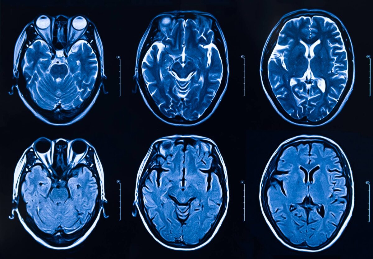
The Long-Term Effects of Surgery on the Brain

Assessment of long-term outcomes is essential in brain surgery. While most studies have traditionally focused on the short-term consequences of brain surgery, more researchers are now reporting long-term outcomes in various cohorts of patients following a variety of invasive neurological interventions [1]. Given the complexity of brain surgery and numerous dangers it poses, healthcare professionals should be knowledgeable about the potential lasting effects of brain surgery so they can provide the best support to recovering patients.  

Before surgery, patients are typically aware of the well-known risks like bleeding and infection, but new research suggests that brain surgery can lead to more lasting issues with critical thinking and memory [2]. In a 2019 study conducted at the University of Wisconsin, researchers found that major neurological surgery is associated with a small and immediate cognitive decline, equivalent to less than five months of aging. The data from the study also suggests that, on average, patients who have brain surgery are nearly twice as likely to experience a decline in cognitive function in the years following their surgeries [3]. However, it is important to note that Dr. Robert Sanders, the senior author of the study, emphasizes that this doubling in risk only affects a small number of patients [3].  

Post-operative cognitive decline is thought to be caused by the immune system’s inflammatory response to surgery [2]. Brain surgery is considered a traumatic event for the body, resulting in the engagement of the immune system and an inflammatory cascade that affects the synaptic plasticity in brain regions involved in learning and memory [2]. If trauma-induced inflammation is not carefully regulated, patients could experience more than a minor cognitive decline [2].  

Even the youngest of patients seem to be susceptible to cognitive decline associated with brain surgery. Over the past 15 years, numerous studies have pointed to an association between commonly used anesthetics and sedatives in surgery and neurodegenerative changes in developing brains [4]. One 2017 study compared the IQ test scores and school grades of two groups of children: those who underwent surgery before the age of four (33,514 children) and those who had no history of surgery (159,619 children) [5]. The results indicate that exposure to surgery with anesthesia before the age of four is linked to lower IQ test scores and poorer academic performance [5].  

Healthcare providers should also be aware that patients who have had brain surgery are at an increased risk for developing depression. In 2008, a study examined the anxiety and depression levels of 72 patients before and after surgery to remove a primary brain tumor [6]. Before brain surgery, 50% of study participants showed general anxiety and 9.7% showed current depression [6]. Anxiety was assessed using the State and Train Anxiety Inventory, and depression was measured using the Zung Self-Rating Depression Scale [6]. Study participants were reassessed at both one month and three months after brain surgery [6]. The data showed no significant change in the percentage of patients with anxiety [6]. However, there was a significant increase in the percentage of patients with depression [6]. These results suggest that a depressive state can develop after brain surgery.  

It is important for healthcare professionals to understand the long-term effects of brain surgery so that recovering patients can be appropriately monitored and supported. Although the lasting effects are dependent both on the extent and location of the operation and the patient profile, healthcare professionals should be attentive to any cognitive or mood changes in patients who have experienced brain surgery.  

References

1. Tellez-Zenteno, J. F., R. Dhar, L. Hernandez-Ronquillo, and S. Wiebe. 2007. “Long-Term Outcomes in Epilepsy Surgery: Antiepileptic Drugs, Mortality, Cognitive and Psychosocial Aspects”. Brain 130 (2): 334-345. doi:10.1093/brain/awl316.  

2. Saxena, Sarah, and Mervyn Maze. 2018. “Impact on The Brain of The Inflammatory Response to Surgery”. La Presse Médicale 47 (4): e73-e81. doi:10.1016/j.lpm.2018.03.011.  

3. Krause, Bryan M, Séverine Sabia, Helen J Manning, Archana Singh-Manoux, and Robert D Sanders. 2019. “Association Between Major Surgical Admissions and The Cognitive Trajectory: 19 Year Follow-Up of Whitehall II Cohort Study”. BMJ, l4466. doi:10.1136/bmj.l4466.  

4. Andropoulos, Dean B. 2017. “Effect of Anesthesia on The Developing Brain: Infant and Fetus”. Fetal Diagnosis and Therapy 43 (1): 1-11. doi:10.1159/000475928.  

5. Glatz, Pia, Rolf H. Sandin, Nancy L. Pedersen, Anna-Karin Bonamy, Lars I. Eriksson, and Fredrik Granath. 2017. “Association of Anesthesia and Surgery During Childhood with Long-Term Academic Performance”. JAMA Pediatrics 171 (1): e163470. doi:10.1001/jamapediatrics.2016.3470.  

6. D’Angelo, Cristina, Antonio Mirijello, Lorenzo Leggio, Anna Ferrulli, Vincenzo Carotenuto, Nadia Icolaro, Antonio Miceli, Vincenzo D’Angelo, Giovanni Gasbarrini, and Giovanni Addolorato. 2008. “State and Trait Anxiety and Depression in Patients with Primary Brain Tumors Before and After Surgery: 1-Year Longitudinal Study”. Journal of Neurosurgery 108 (2): 281-286. doi:10.3171/jns/2008/108/2/0281. 

The Evolution of the Anesthesia Machine

A quick glance around a typical household kitchen reveals a surprising truth: contained therein are the founding mechanisms used to administer anesthesia. The humble beginnings of sponges, towels, and handkerchiefs have evolved into the computerized self-sufficient anesthesia machines used today.

William Morton, a dental surgeon, publicly demonstrated the first successful pain-free surgery using ether anesthesia in 1846. For delivery, he used a glass globe with two valves for the inhalation and diffusion of ether vapor as air circled through. The contraption contained a sponge soaked in ether liquid for expanding the evaporation surface [1]. However, there loomed dangers of uncontrolled ether concentrations and varying amounts of anesthesia required for sedation [2]. Chloroform, a liquid anesthetic, decreased the heart rate more than ether, making it crucial to find a method for controlled administration [3]. By 1847, physician John Snow devised a machine to control vapor pressure using water to stabilize temperature and metal spiraled air passages to saturate ether. With many doctors unwilling to give up simplistic methods and learn complex apparatuses, Snow’s invention of a controlled ether inhaler was largely ignored [4].

Nitrous oxide gas catalyzed a turning point for anesthetics. Its harmless ability to stimulate rather than depress the nervous system and to quickly sedate without irritating the lungs, trumped the risks of ether and chloroform [5]. In 1865, a dental company began manufacturing impractical appliances requiring the immense efforts of holding a wooden mouthpiece while plugging patients’ noses to inhale gas from a bag. Liquid-form nitrous oxide avoided these nuances and began being sold in cylinders attached to a maze of bags, valves, and a mask [6]. To counteract the lack of oxygen in this arrangement, physiologist Paul Bert designed a hyperbaric pressure chamber providing 15% oxygen and 85% nitrous oxide but was unaware of oxygen toxicity at high pressures [7]. Eventually, the dental company’s initial flawed gas bag was redeemed with one of the first representations of an anesthesia machine. With pressure-regulating valves directing gas into a mixing chamber for oxygen and nitrous oxide, patients were safely sedated [4]. A milestone in medicine, anesthesia began transforming into a working machine.

The next century, anesthesia machines faced the challenge of accurately measuring gas concentration and flow. Initially, flow meters were used that relied heavily on the human eye to measure flow pressure based off of bubbling rates of water and moving bobbins. Gradually, rotameters replaced these, allowing medical professionals to self-calculate flow rate from the force of gas exertion. To preserve gas emissions from flow meters, pressure-controlled ventilators were introduced, depending on patients’ lungs to drive pressure and volume flow, but human error still remained [8].

In 1950, Snow’s achievement of temperature stabilization was implemented in new vaporizer designs. Now, delivery of liquid anesthesia depended only on flow rate and temperature. Though “variable bypass vaporizers” eliminated human calculation, they still required manual control to adjust concentrations [4]. With promising results from a computer-controlled anesthesia machine demonstration in 1978, manually operated vaporizers became computer- powered. Monitor feedback directed vaporizers to pump the amount of liquid anesthetic needed for specific vapor concentrations, offloading manual work — and marking the beginning of the twenty-first century.

With corporations releasing different prototypes, hospital workers spoke of “spaghetti syndrome,” as they jumbled up an array of cords from various machines [9]. Responding to these outcries, companies introduced monitors, anesthesia machines, and ventilators in one complete set to create the anesthesia workstation. From sponges to computers, the evolution of the anesthesia machine has now culminated in a single, portable, computerized station. Ideally, future evolutions will continue to safeguard the balance between computer and human responsibilities.

References

[1] Duncum B. (1994). The Development of Inhalation Anaesthesia. Royal Society of Medicine Press Ltd, London, 553-9, 106-8

[2] Bigelow, H. J. (1846). Insensibility during Surgical Operations Produced by Inhalation. The Boston Medical and Surgical Journal35(16), 309–317. https://doi.org/10.1056/nejm184611180351601

[3] J. Y. Simpson. (1958). New Anaesthetic Agent as a Substitute for Sulphuric Ether in Surgery and Midwifery, BJA: British Journal of Anaesthesia, Volume 30, Issue 11. 545 550, https://doi.org/10.1093/bja/30.11.545

[4] Eger, E. I., Saidman, L. J., & Westhorpe, R. N. (2014). Anesthesia Machines and Breathing Systems: An Evolutionary Success Story. The Wondrous Story of Anesthesia, 9781461484417, 703–714. https://doi.org/10.1007/978-1-4614-8441-7

[5] Weyde, V. (1864). Nitrous Oxide Gas. The New York Times. Retrieved from https://www.nytimes.com/1864/05/28/archives/nitrous-oxide-gas.html

[6] Thomas, K. B. (1976). The Development of Anaesthetic Apparatus. The 2 Oxide Series45(1), 74-103. https://doi.org/10.1097/00000542-197607000-00030

[7] Neuman, T. (2008). Physiology and medicine of hyperbaric oxygen therapy. Philadelphia, PA: Saunders Elsevier. 7-9

[8] Dorsch, J. A., & Dorsch, S. E. (2012). Understanding anesthesia equipment: Fifth editionUnderstanding Anesthesia Equipment: Fifth Edition. 1–1056. Wolters Kluwer Health Adis (ESP).

[9] Westhorpe, R. N. (1992). The anesthetic machine in the 1990s. Anesthesiology Review. 46-55.

Anesthetic Management of Patient with Obstructive Sleep Apnea

Safe anesthetic management of patients at risk for or who carry a diagnosis of obstructive sleep apnea (OSA) is imperative to guiding this subset of patients through the perioperative period. OSA carries perioperative risk both due to the direct respiratory effects of the disease, comorbidities of patients with OSA, as well as the associated long-term sequelae of patients with untreated or severe OSA. There is a relatively low number of studies examining best practices in anesthesia regarding patients with OSA, so much of the literature and recommendations focus on small studies and expert opinion. 

OSA is a disorder where upper airway tissue collapses into the upper airway during sleep, causing periods of apnea or hypopnea that often requires the patient (unbeknownst to them) to wake briefly to catch their breath. It is a quite common disorder, with up to 15-30% of males and 10-15% of females in North America carrying the diagnosis. Risk factors include male gender, obesity, smoking, craniofacial abnormalities, and older age. 

The first step for an anesthesia provider in the pre-operative period is to ascertain whether a patient carries a diagnosis of OSA, and if they do not, if they have risk factors for the disorder. If the patient carries an OSA diagnosis and has therefore had a sleep study (which is the gold standard for diagnosis), then the results of that study should be read by the anesthesia provider. Severity of OSA is classified by the apnea-hypopnea index (AHI), which is a scale based on the number of apneic or hypopneic episodes per hour of sleep. Less than 5 events indicates no OSA, 5-15 events is mild, 15-30 is moderate, and greater than 30 is severe. The more severe the disease, the more at risk a patient is both for perioperative morbidity, as well as long-term sequelae of the disease. If a patient is not diagnosed with OSA, there are a number of assessments that can be used to indicate risk. The best combination of sensitivity and specificity is the STOP-BANG questionnaire, which includes a variety of questions (snoring, daytime sleepiness, observed awakenings sleep partner, etc.). There are seven total questions, and a confirmatory answer to three or more questions indicates a high risk of OSA. 

The most important aspect of an anesthesia provider’s pre-operative assessment and perioperative plan is being aware of the risks factors associated with long-standing or untreated OSA; these include hypertension, coronary artery disease, heart failure, arrythmias, and stroke. Patients with OSA and pulmonary disease are also at particular risk for pulmonary hypertension. As a result, in a patient with OSA, functional status is an essential part of the preoperative evaluation, and cardiac catheterization or transthoracic echocardiography may be indicated prior to surgery as well.  

Once a pre-operative assessment is complete, OSA patients have a higher risk of a number of intraoperative difficulties. OSA patients are often obese, and as a result can be difficult intravenous access, high aspiration risk, and can have difficult airways, with a particularly high risk of being difficult to mask ventilate. As a result, avoiding general anesthesia using regional techniques can and should be used when possible. However, even mild to moderate amounts of sedation can be risky in OSA patients, as they by definition have airways that obstruct quite easily. When general anesthesia is required, an RSI may be indicated depending on the patient’s weight and comorbidities, and in other circumstances an awake fiberoptic intubation may be the safest approach. 

Post-operatively, hypoventilation is the most important concern for OSA patients. Patients with OSA tend to be sensitive to narcotics, and as stated above, obstruct their airways easily. This combination can be devastating if high doses of narcotics are used, so this should be avoided during surgery and afterwards. In addition, moderate to severe OSA patients are not good candidates for same day surgery, and should be monitored in a hospital setting postoperatively until the effects of anesthesia have completely worn off. The use of CPAP in OSA patients post-operatively has been studied without clear conclusions, but positive pressure ventilation techniques should, at the least, be readily available to post-operative providers. 

References: 

Chung et al. Society of Anesthesia and Sleep Medicine Guidelines on Preoperative Screening and Assessment of Adult Patients With Obstructive Sleep Apnea. Anesth Analg. 2016 Aug;123(2):452-73. doi: 10.1213/ANE.0000000000001416. 

Okoronkwo U. Ogan, M.D.; David J. Plevak, M.D. Anesthesia Safety Always an Issue with Obstructive Sleep Apnea. APSF Newsletter. Volume 12, No. 2, 1997. 

Young T, Palta M, Dempsey J, et al. The occurrence of sleep-disordered breathing among middle-aged adults. N Engl J Med 1993; 328:1230. 

Sunwoo JS, Hwangbo Y, Kim WJ, et al. Prevalence, sleep characteristics, and comorbidities in a population at high risk for obstructive sleep apnea: A nationwide questionnaire study in South Korea. PLoS One 2018; 13:e0193549. 

Mason M, Cates CJ, Smith I. Effects of opioid, hypnotic and sedating medications on sleep-disordered breathing in adults with obstructive sleep apnoea. Cochrane Database Syst Rev 2015; :CD011090. 

Oxytocin Administration for Cesarean Delivery

Hormonal processes of childbearing are essential to pregnancy, labor, birth, breastfeeding and the maternal–child attachment.1 Hormone release in both the mother and the baby is coordinated to optimize outcomes, such as maternal and fetal readiness for labor.1 Oxytocin is a hormone that is produced naturally by the body, but also administered to induce labor that may not have started on its own2 or to prevent postpartum blood loss.3 To provide the best care to their parturient patients, anesthesia providers should be familiar with the uses of oxytocin as well as protocols for oxytocin in Cesarean delivery.4 

Prelabor preparation in the maternal body involves many complex hormonal and physiological changes.1 These changes, which occur in the weeks, days and hours before the onset of labor in humans and animals, include rising estrogen levels, activating the uterus for efficient labor; increases in oxytocin and prostaglandin activity, causing cervical ripening; increased inflammation, which activates the cervix and uterus; increased uterine oxytocin receptors, allowing for effective contractions during labor and reduced bleeding after birth; increased central nervous system (CNS) receptors for beta-endorphins, contributing to endogenous analgesia in labor; and elevations in mammary and CNS oxytocin and prolactin receptors, which promote breastfeeding and maternal-infant attachment after birth.1 Similarly, changes in hormones and neurotransmitters affect the baby’s adaptations for labor as well.1 In some situations, such as scheduled birth, endogenous hormonal activities do not align with external needs. In these cases, clinicians will administer exogenous hormones to encourage certain processes. Once the cervix has been softened using prostaglandins, a health professional will provide a synthetic version of oxytocin (known as Pitocin) to cause uterine contraction and thus induce labor.5 Oxytocin is more effective at speeding up labor that has already begun than it is at beginning the labor process.5 When epidural analgesia is used during vaginal birth, the resulting reduction in maternal oxytocin may require the clinician to administer exogenous oxytocin to compensate.1 Also, clinicians commonly administer oxytocin after both vaginal and operative delivery to initiate and maintain adequate uterine contractility, thus minimizing blood loss and preventing postpartum hemorrhage.3 Evidently, oxytocin and various other hormones are crucial to efficient labor, and may be administered by clinicians to provoke certain essential processes. Future research is needed to assess the long-term effects of exogenous oxytocin exposure during labor and delivery.6 

Anesthesia providers are highly involved in Cesarean sections, and one of their many roles can involve oxytocin administration. Because Cesarean section is fetal birth through incisions in the abdominal wall and uterine wall, it does not involve the same endogenous hormone releases as does vaginal birth.7 Thus, oxytocin infusion at Cesarean section may help maintain uterine contractility throughout the procedure and immediately postpartum, when most hemorrhage (blood loss) occurs.7 The protocols surrounding oxytocin administration for Cesarean section are complicated and must be followed precisely.4 However, there remains a lack of consensus among anesthesiology practitioners regarding optimal dose and mode of administration.8 For example, Terblanche et al.’s review showed no conclusion for or against the effectiveness of 5IU oxytocin versus a lower dose for combatting postpartum blood loss.8 However, Balki et al. found that women requiring Cesarean delivery for labor arrest required approximately 3IU of intravenous oxytocin to achieve effective uterine contraction after delivery.9 Kovacheva et al. found that when compared to women receiving continuous infusions of oxytocin, women who received a low-dose bolus of 3IU oxytocin achieved the same adequate uterine tone while using less oxytocin.10 On the other hand, high doses of oxytocin can cause cardiovascular issues or desensitization of oxytocin receptors.11 Some researchers show that timing may be an important factor in oxytocin infusion. Bhattacharya et al. found that in elective Cesarean delivery patients, bolus oxytocin of 3IU over 15 seconds and infusion of oxytocin at 3IU over five minutes had comparable uterotonic effects, but the bolus administration resulted in significantly more cardiovascular events.3 Additionally, Foley et al.’s retrospective chart review found that pre-Cesarean exposure to oxytocin led patients to require a high postpartum oxytocin infusion rate more often than patients who were not exposed.12 Clinicians have yet to determine the optimal dose and timing of oxytocin doses in Cesarean section.11 

Anesthesia providers often care for parturient patients who are experiencing various hormonal changes. Sometimes, vaginal or Cesarean delivery requires exogenous oxytocin to induce labor or prevent postpartum blood loss. Future studies should evaluate the optimal dose of oxytocin doses, the most effective mode of administration and the possible long-term implications of oxytocin administration during Cesarean delivery.  

1.Buckley SJ. Executive Summary of Hormonal Physiology of Childbearing: Evidence and Implications for Women, Babies, and Maternity Care. The Journal of Perinatal Education. 2015;24(3):145–153. 

2.Hormone Health Network. What is Oxytocin? Glands & Hormones A–Z November 2018; https://www.hormone.org/your-health-and-hormones/glands-and-hormones-a-to-z/hormones/oxytocin

3.Bhattacharya S, Ghosh S, Ray D, Mallik S, Laha A. Oxytocin administration during cesarean delivery: Randomized controlled trial to compare intravenous bolus with intravenous infusion regimen. Journal of Anaesthesiology, Clinical Pharmacology. 2013;29(1):32–35. 

4.Balki M, Tsen L. Oxytocin protocols for Cesarean delivery. International Anesthesiology Clinics. 2014;52(2):48–66. 

5.Mayo Clinic. Labor induction. Tests & Procedures September 11, 2017; https://www.mayoclinic.org/tests-procedures/labor-induction/about/pac-20385141

6.Erickson EN, Emeis CL. Breastfeeding Outcomes After Oxytocin Use During Childbirth: An Integrative Review. Journal of Midwifery & Women’s Health. 2017;62(4):397–417. 

7.Abbas AM. Oxytocin Administration During Cesarean Section. ClinicalTrials.gov: U.S. National Library of Medicine; April 23, 2018. 

8.Terblanche NCS, Picone DS, Otahal P, Sharman JE. Paucity of evidence for the effectiveness of prophylactic low-dose oxytocin protocols (<5 IU) compared with 5 IU in women undergoing elective caesarean section: A systematic review of randomised controlled trials. European Journal of Anaesthesiology. 2018;35(12):987–989. 

9.Balki M, Ronayne M, Davies S, et al. Minimum Oxytocin Dose Requirement After Cesarean Delivery for Labor Arrest. Obstetrics & Gynecology. 2006;107(1):45–50. 

10.Kovacheva VP, Soens MA, Tsen LC. A Randomized, Double-blinded Trial of a “Rule of Threes” Algorithm versus Continuous Infusion of Oxytocin during Elective Cesarean Delivery. Anesthesiology: The Journal of the American Society of Anesthesiologists. 2015;123(1):92–100. 

11.Yamaguchi ET, Siaulys MM, Torres MLA. Oxytocin in cesarean-sections. What’s new? Brazilian Journal of Anesthesiology (English Edition). 2016;66(4):402–407. 

12.Foley A, Gunter A, Nunes KJ, Shahul S, Scavone BM. Patients Undergoing Cesarean Delivery After Exposure to Oxytocin During Labor Require Higher Postpartum Oxytocin Doses. Anesthesia and Analgesia. 2018;126(3):920–924.