Spirituality in Medicine

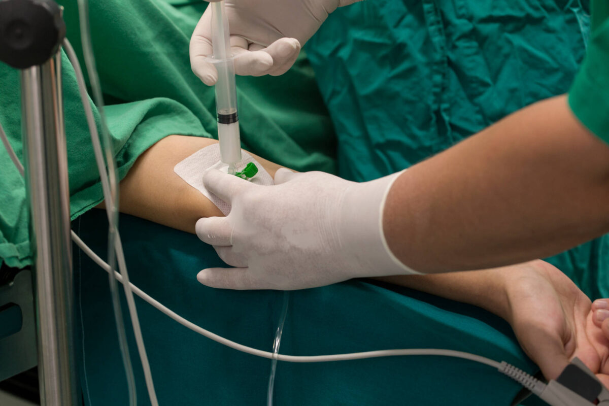
Spirituality is an important part of medical care that many patients desire. Patients facing major illnesses or injuries are frequently in once-in-a-lifetime situations that they could not have emotionally prepared for and often turn to religion and spirituality as a source of comfort. Religion and spirituality are associated with positive medical and psychiatric outcomes, especially in older adults, which makes it an important part of caring for the patient as a whole in medicine. There has been much published data on how religion and spirituality can improve depression, anxiety, and substance use disorders.1 While hospitals frequently have chaplains for these discussions, many patients hope that physicians can engage in these conversations as well. In a study with hospital inpatients, 77% of the inpatients said physicians should consider their spiritual needs as it relates to their health, yet 68% of patients said their physician never discussed religious beliefs with them.2 This suggests there is a gap between what patients hope to receive from medicine in terms of discussions on spirituality and what physicians are delivering.

In a study investigating the medical community’s opinion of religion and spirituality, a survey with almost 900 resident physician respondents from all clinical specialties demonstrated that 75% of residents believed religion and spirituality was important for patient health and that it was important to discuss with patients.3 However, only 14.4% of residents reported doing so in practice. Reasons for why they did not included concern for maintaining professional neutrality and concern about offending patients. Interestingly, in another study looking at how physicians’ beliefs influence their frequency of religious and spiritual discussions, physicians who self-reported to be religious or spiritual had these discussions with patients more than non-religious/spiritual physicians did.4 This suggests physicians who are non-religious/spiritual may be less comfortable initiating these conversations and are at a disadvantage in providing religious and spiritual support to patients. In addition, providers with diverse beliefs can help the field as a whole better address patients’ needs.

The American Association of Medical Colleges (AAMC) publishes objectives for medical students, which includes the ability to ask about spiritual history and understand how spirituality can improve healthcare outcomes.5 Because spirituality can affect the patient’s health, all providers should be educated in how to have these discussions in the context of medicine regardless of their own personal beliefs. However, in practice, medical schools and residencies train learners very little in the skills needed to be comfortable with these discussions. There is little to no formal training or practice which would likely increase physicians’ comfort in asking about religion and spirituality. Increased training is necessary in the future to have more holistic medical care that takes into account the religious and spiritual preferences of the patient, in the hopes of improving their healthcare outcomes.

References

  1. Koenig HG. Religious attitudes and practices of hospitalized medically ill older adults. Int J Geriatr Psychiatry. 1998 Apr;13(4):213-24. doi: 10.1002/(sici)1099-1166(199804)13:4<213::aid-gps755>3.0.co;2-5. PMID: 9646148.
  2. King DE, Bushwick B. Beliefs and attitudes of hospital inpatients about faith healing and prayer. J Fam Pract. 1994 Oct;39(4):349-52. PMID: 7931113.
  3. Vasconcelos APSL, Lucchetti ALG, Cavalcanti APR, da Silva Conde SRS, Gonçalves LM, do Nascimento FR, Chazan ACS, Tavares RLC, da Silva Ezequiel O, Lucchetti G. Religiosity and Spirituality of Resident Physicians and Implications for Clinical Practice-the SBRAMER Multicenter Study. J Gen Intern Med. 2020 Dec;35(12):3613-3619. doi: 10.1007/s11606-020-06145-x. Epub 2020 Aug 19. PMID: 32815055; PMCID: PMC7728988.
  4. Franzen AB. Influence of Physicians’ Beliefs on Propensity to Include Religion/Spirituality in Patient Interactions. J Relig Health. 2018 Aug;57(4):1581-1597. doi: 10.1007/s10943-018-0638-7. PMID: 29876717.
  5. Association of American Medical Colleges (AAMC). Contemporary issues in medicine: communication in medicine. Medical school objectives project, Report III. 1999

Laryngeal Mask Airway Removal

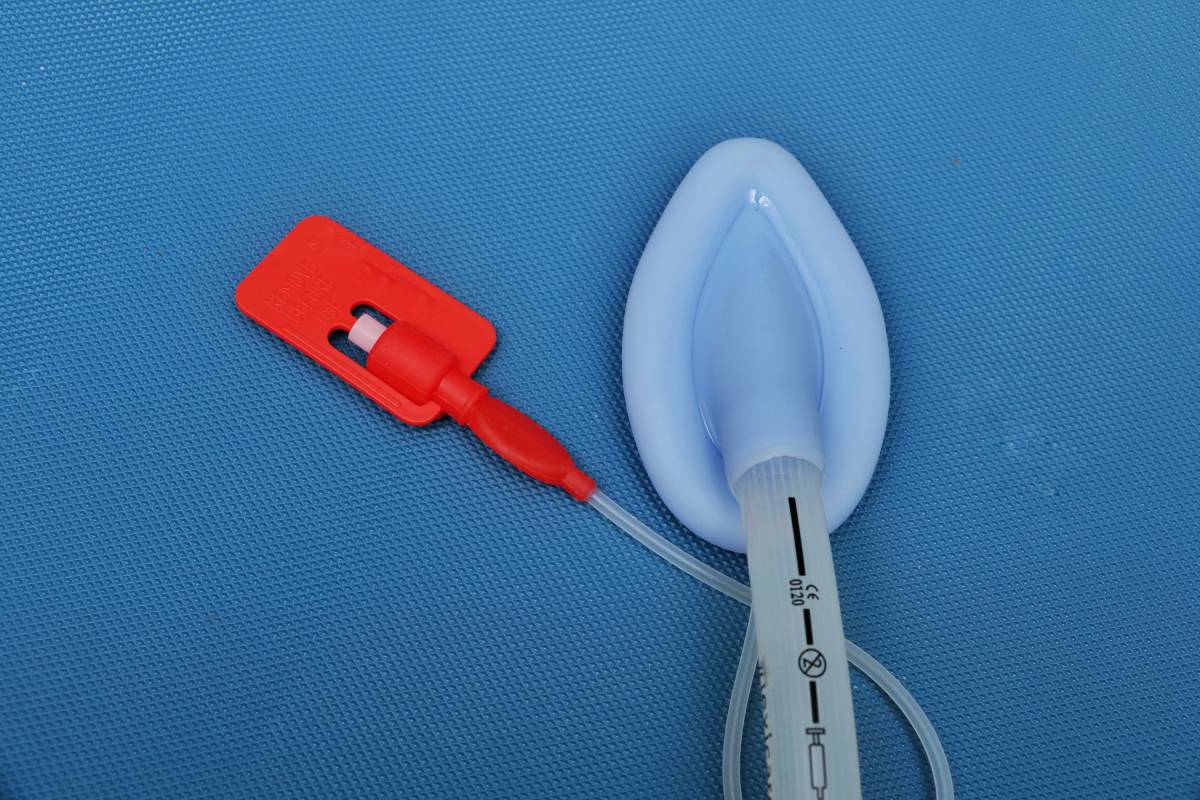
Laryngeal mask airway (LMA) facilitates surgeries by providing high airtightness, low stimulation, and easier management of the supraglottic airway [1]. It is widely used to control the airways of pediatric surgical patients [2]. Unfortunately, laryngeal mask airway removal can result in adverse airway events, so anesthetic choice and timing must be carefully considered to prevent such occurrences [1].

Generally, isoflurane and sevoflurane are the anesthetic agents of choice for LMA [3]. This is because they tend to promote faster recovery [3]. However, other agents could reduce the risk of adverse airway-related events and thus merit consideration for use alongside LMA. One such agent is desflurane. Although desflurane does not permit as fast a recovery as other inhaled anesthetic agents, its pharmacokinetic qualities and rapid metabolism can render it appropriate for use in low-flow systems [3]. To address desflurane’s longer laryngeal mask airway removal times, medical teams can administer it in an entropy-guided low-flow context [3]. This technique has demonstrably promoted lower consumption of anesthetics and shorter removal time compared with minimal alveolar concentration-guided anesthesia [3].

Other variations on anesthetic practice that promote better outcomes include the combined use of propofol with isoflurane, or dexmedetomidine with sevoflurane [2, 4]. Kumar and colleagues found that deep anesthesia via a propofol-isoflurane combination resulted in a lower incidence of teeth clenching and airway obstruction, and a shorter emergence duration compared to isoflurane alone [2]. On the other hand, Bhat and colleagues found that dexmedetomidine (1 µg /kg) with sevoflurane decreased emergence agitation and promoted smoother removal of LMA than normal saline or lower dose dexmedetomidine (0.5 1 µg /kg) with sevoflurane [4].

As for timing, physicians looking to remove a patient’s laryngeal mask airway generally contemplate two options: removal during deep anesthesia, or removal following emergence [2]. Significant challenges are associated with each possibility. Removal under deep anesthesia risks events such as glossocoma and pharyngalgia, while removal in the awake state can lead to PACU complications and coughing [1].

Several experiments have sought to determine which option is preferable in different contexts. Sun et al conducted a randomized controlled trial consisting of children undergoing squint correction operations with general anesthesia [1]. They measured adverse airway events such as glossocoma, pharyngalgia, coughing, and laryngeal spasm rates [1]. When lidocaine cream was applied to the LMA cuff prior to insertion, removal in the awake state was generally preferable to deep anesthesia removal [1]. This was also true when hydrosoluble lubricant had been applied to the LMA instead of lidocaine cream [1].

Ramgolam et al found that tonsillectomy patients aged 0 to 16 years old exhibited no significant difference in adverse events overall, regardless of whether removal occurred while they were under deep anesthesia or awake [5]. However, subjects who experienced deep removal were more likely to suffer respiratory adverse events [5]. In a larger pool of subjects, this could mean that deep anesthesia laryngeal mask airway removal results in increased costs, hospital stays, and postsurgical adverse events [6].

On a parallel note, Asahi and colleagues narrowed their focus to pediatric patients with special needs [7]. These patients require extra consideration, given their difficulty complying with instructions during removal [7]. The 80 patients were divided into two groups: removal during the pre-awake state and the deep anesthesia state [7]. The pre-awake states exhibited gross body movement, more clenching, and difficult ventilation, suggesting that deep anesthesia may be preferable for this subsection of patients [7].

The optimal LMA removal technique requires a complex inquiry into anesthetic agents, timing, and patients’ individual abilities to comply with instructions. By accounting for these factors, physicians can provide for easier removal and fewer adverse airway events.

References 

[1] R. Sun et al., “The impact of topical lidocaine and timing of LMA removal on the incidence of airway events during the recovery period in children: a randomized controlled trial,” BMC Anesthesiology, vol. 21, no. 10, p. 410-417, January 2021. [Online]. Available: https://doi.org/10.1186/s12871-021-01235-7

[2] D. Kumar et al., “Isoflurane alone versus small dose propofol with isoflurane for removal of laryngeal mask airway in children-a randomized controlled trial,” Journal of Pakistan Medical Association, vol. 69, no. 11, p. 1596-1600, November 2019. [Online]. Available: https://doi.org/10.1186/s12871-021-01235-7

[3] S. Mishra et al., “Effect of entropy-guided low-flow desflurane anaesthesia on laryngeal mask airway removal time in children undergoing elective ophthalmic surgery – A prospective, randomised, comparative study,” Indian Journal of Anaesthesia, vol. 63, no. 6, p. 485-490, June 2019. [Online]. Available: http://doi.org/10.4103/ija.IJA_237_19

[4] R. Bhat et al., “Study of dose related effects of dexmedetomidine on laryngeal mask airway removal in children -a double blind randomized study,” Anaesthesia, Pain & Intensive Care, vol. 22, no. 3, p. 368-373, July-September 2018. [Online]. Available: https://apicareonline.com/index.php/APIC/article/view/74

[5] A. Ramgolam et al., “Deep or awake removal of laryngeal mask airway in children at risk of respiratory adverse events undergoing tonsillectomy—a randomised controlled trial,” British Journal of Anaesthesia, vol. 120, no. 3, p. 571-580, March 2018. [Online]. Available: https://doi.org/10.1016/j.bja.2017.11.094

[6] M. Oofuvong et al., “Excess costs and length of hospital stay attributable to perioperative respiratory events in children,” Anesthesia and Analgesia, vol. 120, no. 2, p. 411-419, February 2015. [Online]. Available: https://doi.org/10.1213/ANE.0000000000000557

[7] Y. Asahi et al., “Excess costs and length of hospital stay attributable to perioperative respiratory events in children,” Journal of Oral and Maxillofacial Surgery, Medicine, and Pathology, October 2021. [Online]. Available: https://doi.org/10.1016/j.ajoms.2021.09.004

Workplace Mistreatment in Medicine

Workplace mistreatment is a widespread issue, occurring in all subspecialties in medicine and at all levels of the medical hierarchy. Mistreatment includes discrimination, verbal or physical abuse, and sexual harassment. More subtle forms may include micro-aggressions or gaslighting arising from conscious or unconscious bias. Mistreatment toward medical professionals may lead to burnout, which is defined by the World Health Organization as emotional exhaustion and fatigue. Burnout can contribute to depression and suicidal ideation, which increases the risk for suicide completion. 28% of resident physicians experience a major depressive episode during training compared to 7-8% of similarly aged adults in the U.S. general population1. An estimated 300 physicians die by suicide every year at nearly twice the rate of the general population1. Not only is physician mistreatment detrimental to the physicians, but it also negatively impacts patient care. Long-term effects cause suboptimal care practices, medical and medication errors, and decreased patient satisfaction with medical care5. It is important to understand the causes and components leading to these statistics in order to prevent them in the future.

Among resident physicians, commonly reported examples of workplace mistreatment include gender discrimination, racial discrimination, verbal abuse and sexual harrassment.2 In a 2020 meta-analysis, researchers found 64.1% of resident physicians experienced some form of intimidation, harassment, or discrimination.3 The most common forms of mistreatment were verbal, physical, and sexual abuse. The people who most frequently caused the mistreatment were relatives or friends of patients, nurses, and patients. In another study, researchers found similar results with the additional conclusion that women experienced more gender discrimination and sexual harassment. Patients and their families more often discriminated by gender while attending physicians were reported more for sexual harassment and abuse.2

Even after completion of residency training, attending physicians can experience workplace mistreatment. A 2021 cross-sectional study surveyed nearly 600 attending anesthesiologists about microaggressions. 94% of female physicians reported experiencing sexist microaggressions of hearing or seeing degrading female terms and images. 81% of minority physicians experienced racial and ethnic microaggressions. Of note, levels of burnout were higher among female and minority physicians, associating increased workplace mistreatment with increased levels of burnout.

One way to combat workplace mistreatment in medicine is to educate all levels of healthcare workers about what abuse looks like and empower them to be “upstanders” instead of passive bystanders.5 Strategies include educating healthcare workers on how to intervene when they observe abuse or mistreatment, either through direct or indirect intervention. Direct intervention involves acknowledging abuse or micro-aggressions and starting a conversation about it such as saying, “I heard you say this, which makes me feel…”. By addressing the behavior, the perpetrator may realize the behavior is unacceptable in the workplace and can self-reflect on the perspective that led them to behave in that manner. Indirect intervention redirects attention by shifting the conversation to other topics, which may be a valuable tool when bystanders do not feel comfortable directly addressing the perpetrator. With widespread education, time, and cultural change, hopefully, the prevalence of workplace mistreatment will decrease, leading to downstream effects of better mental health for medical professionals in the future.

References

  1. American Foundation for Suicide Prevention. (n.d.). 10 Facts About Physician Suicide and Mental Health [Brochure]. New York, NY: Author.
  2. Mata DA, Ramos MA, Bansal N, Khan R, Guille C, Angelantonio ED, Sen S. (2015). Prevalence of Depression and Depressive Symptoms among Resident Physicians. JAMA, 314(22), 2373. doi:10.1001/jama.2015.15845. PMID: 26647259; PMCID: PMC4866499.
  3. Bahji A, Altomare J. Prevalence of intimidation, harassment, and discrimination among resident physicians: a systematic review and meta-analysis. Can Med Educ J. 2020 Mar 16;11(1):e97-e123. doi: 10.36834/cmej.57019. PMID: 32215147; PMCID: PMC7082478.
  4. Sudol NT, Guaderrama NM, Honsberger P, Weiss J, Li Q, Whitcomb EL. Prevalence and Nature of Sexist and Racial/Ethnic Microaggressions Against Surgeons and Anesthesiologists. JAMA Surg. 2021 May 1;156(5):e210265. doi: 10.1001/jamasurg.2021.0265. Epub 2021 May 12. PMID: 33760000; PMCID: PMC7992024.
  5. Ehie O, Muse I, Hill L, Bastien A. Professionalism: microaggression in the healthcare setting. Curr Opin Anaesthesiol. 2021 Apr 1;34(2):131-136. doi: 10.1097/ACO.0000000000000966. PMID: 33630771; PMCID: PMC7984763.

Intranasal Steroids and COVID-19

SARS-CoV-2, the virus underlying the Covid-19 pandemic, is known to cause a host of symptoms, including fever, cough, dyspnea, sputum production, myalgia, arthralgia, headache, gastrointestinal issues, rhinorrhea, sore throat, and loss of olfactory ability/taste. Olfactory symptoms are common among Covid-19 patients; in fact, one study found that 86 percent of infected individuals experienced detectable olfactory symptoms.1 Given the prevalence of the virus, it has been of heightened interest to find methods of sino-nasal symptom alleviation. One recently proposed method includes the use of nasal steroids to treat Covid-19.

Nasal steroid sprays are traditionally used to alleviate allergy or, less commonly, non-allergy related inflammation in the nasal cavity. The steroid is suspended within pressurized gas, allowing for dispersion into the nasal cavity upon release from the canister. The effect of the steroid is a reduction in the size of blood vessels and surrounding tissue, therefore treating acute congestion and making respiration easier.

Several studies seem to suggest that chronic use of intranasal steroids can help alleviate some of the olfactory symptoms which characterize Covid-19. A study published by İşlek et al., titled, “Evaluation of effects of chronic nasal steroid use on rhinological symptoms of COVID-19 with SNOT-22 questionnaire,” showed that Covid-19 positive patients who used intranasal mometasone furoate spray once in a day experienced milder olfactory symptoms and had shorter recovery times when it came to post-viral olfactory dysfunction.2 Notably, symptoms were not prevented, merely alleviated. However, another slightly contradictory study found that anosmia – one of the most prevalent and characteristic symptoms of Covid-19 – was not alleviated in patients who self-administered intranasal steroids daily for three weeks.3 Alternatively, some researchers have hypothesized that nasal steroids could be used in the fight against Covid-19 infection itself, as opposed to merely the symptoms. It has been shown that SARS-CoV-2 utilizes the host cell’s ACE2 receptor as a point of entry and adhesion.4 Given their vital role in regulating blood pressure, wound healing, and inflammation, ACE2 receptors can be found in most cell types throughout the body; however, they are expressed in particularly high levels in epithelial tissue and nasal mucosa.5 In fact, studies have shown that higher viral load of SARS-CoV-2 can be found in nasal swabs as compared to throat swabs, a difference which has been attributed to higher levels of ACE2 expression in the nasal mucosa.6 Conversely, research has demonstrated that ACE2 expression in nasal epithelial cells in vitro is significantly suppressed by dexamethasone.5 Taken together, these findings point towards the possibility that corticosteroids could prevent viral infection and replication in nasal epithelial cells by reducing potential entry points into cells. However, transmission is possible through a number of different avenues, so any protection conferred by nasal corticosteroids would be inherently limited.

In conclusion, the use of nasal corticosteroids is a promising approach for the treatment of Covid-19 nasal symptoms, particularly congestion. However, more remains to be learned as to whether there is a possibility of infection prevention, and the literature on anosmia remains inconsistent. We will likely continue to learn more about intranasal corticosteroids and Covid-19 throughout the course of the pandemic.

References 

1 Lechien, J. R., Chiesa-Estomba, C. M., De Siati, D. R., Horoi, M., Le Bon, S. D., Rodriguez, A., Dequanter, D., Blecic, S., El Afia, F., Distinguin, L., Chekkoury-Idrissi, Y., Hans, S., Delgado, I. L., Calvo-Henriquez, C., Lavigne, P., Falanga, C., Barillari, M. R., Cammaroto, G., Khalife, M., Leich, P., … Saussez, S. (2020). Olfactory and gustatory dysfunctions as a clinical presentation of mild-to-moderate forms of the coronavirus disease (COVID-19): a multicenter European study. European archives of Oto-rhino-laryngology, 277(8), 2251–2261. https://doi.org/10.1007/s00405-020-05965-1 

2 İşlek, A., & Balcı, M. K. (2021). Evaluation of effects of chronic nasal steroid use on rhinological symptoms of COVID-19 with SNOT-22 questionnaire. Pharmacological Reports, 73(3), 781–785. https://doi.org/10.1007/s43440-021-00235-1 

3 Abdelalim, A. A., Mohamady, A. A., Elsayed, R. A., Elawady, M. A., & Ghallab, A. F. (2021). Corticosteroid nasal spray for recovery of smell sensation in COVID-19 patients: A randomized controlled trial. American Journal of Otolaryngology, 42(2), 102884. https://doi.org/10.1016/j.amjoto.2020.102884 

4 Hoffmann, M., Kleine-Weber, H., Schroeder, S., Krüger, N., Herrler, T., Erichsen, S., Schiergens, T. S., Herrler, G., Wu, N. H., Nitsche, A., Müller, M. A., Drosten, C., & Pöhlmann, S. (2020). SARS-CoV-2 Cell Entry Depends on ACE2 and TMPRSS2 and Is Blocked by a Clinically Proven Protease Inhibitor. Cell, 181(2), 271–280.e8. https://doi.org/10.1016/j.cell.2020.02.052 

5 Saheb Sharif-Askari, F., Saheb Sharif-Askari, N., Goel, S., Fakhri, S., Al-Muhsen, S., Hamid, Q., & Halwani, R. (2020). Are patients with chronic rhinosinusitis with nasal polyps at a decreased risk of COVID-19 infection?. International Forum of Allergy & Rhinology, 10(10), 1182–1185. https://doi.org/10.1002/alr.22672 

6 Zou, L., Ruan, F., Huang, M., Liang, L., Huang, H., Hong, Z., Yu, J., Kang, M., Song, Y., Xia, J., Guo, Q., Song, T., He, J., Yen, H. L., Peiris, M., & Wu, J. (2020). SARS-CoV-2 Viral Load in Upper Respiratory Specimens of Infected Patients. The New England Journal of Medicine, 382(12), 1177–1179. https://doi.org/10.1056/NEJMc2001737 

Ketofol: Combination Propofol and Ketamine

Despite their demonstrated efficacy as sedation and analgesics, propofol and ketamine can result in adverse events [1]. Propofol can lead to hypotension, respiratory depression, hypoventilation, and loss of airway reflexes [2]. Conversely, ketamine does not risk the compromise of a patient’s airway reflexes, but it can cause hypertension, emergence delirium, vomiting, and tachycardia [2]. Ketamine can also impede the central nervous system [1]. To avoid the complications associated with each medication, researchers developed ‘ketofol’ as a combination of propofol and ketamine [1].

At first glance, ketofol appeared to be an effective replacement for ketamine and propofol. In 1999, Friedberg conducted a large meta-analysis of 2,059 operations performed on 1,264 patients anesthetized using combination propofol and ketamine [3]. None of the patients reported hallucinations, uncontrolled pain, vomiting, and nausea, and all of them were satisfied with their anesthetic regimen [3]. The study concluded that ketofol is a safe and effective anesthetic [3]. More recently, Slavik and Zed retrospectively analyzed 23 studies, all of which were centered on the efficacy of propofol and ketamine in combination [1]. They concluded that there was not substantial literature to demonstrate that ketofol evades the adverse cardiovascular events associated with ketamine and propofol [1]. Furthermore, ketamine-propofol did not appear to provide better analgesia than propofol alone [1]. While these findings do not necessarily contradict Friedberg’s conclusion, they certainly demonstrate the need for more research on ketofol.

For anesthesia providers considering using ketofol during an operation, knowledge of the appropriate ratio to administer is essential. Several studies have compared different ratios among themselves, as well as to anesthetic monotherapies, such as propofol alone [4]. One such study drew a comparison between 2:1, 3:1, and 4:1 propofol-ketamine combinations; propofol alone; and a propofol-fentanyl combination [4]. All of the ketofol mixtures provided patients with effective analgesia [4]. However, the 3:1 and 4:1 mixtures led to especially quick recovery and discharge times [4]. Additionally, the 4:1 mixture was responsible for the lowest incidences of postoperative dizziness and respiratory depression among the propofol-ketamine mixtures [4]. While these are the results of only one experiment, they could help guide physicians toward the ideal ratio with which to create ketofol.

Despite its uncertain benefits, ketofol has already found meaningful application in hospital emergency departments (ED). Phillips et al. studied the efficacy of combination ketamine and propofol in an ED during brief procedures, such as fracture manipulation [5]. Of the 28 patients studied, none required intervention or underwent respiratory depression [5]. Compared to the propofol group, the ketofol group was closer to their target bispectral index monitoring score and experienced less hypotension [5]. Willman and Andolfatto also encountered promising results when analyzing the cases of 114 patients who received ketofol in an ED [6]. No patient experienced vomiting or hypotension, and 96.5% of patients did not require additional sedatives [6]. Together, these results suggest there may be cardiovascular benefits, among others, to the use of ketofol as a sedative.

Still, ketofol does not seem so promising in all contexts. One such context is pediatrics. A recent meta-analysis of 29 studies sought to determine the efficacy of ketofol in operations conducted on children [7]. Hayes et al. did identify reduction of some risks due to ketofol–including apnea, hypotension, and bradycardia [7]. Yet, they also found that ketofol may increase children’s risk of respiratory adverse events, hypertension, and tachycardia [7]. Another study did not find a significant difference in sedative quality between ketofol and ketamine, with adverse events occurring at comparable frequencies [2]. Whether these results are the symptoms of imperfect ratios, or indicative of a greater failure on the part of ketofol, warrants continued investigations into the mechanisms of combination propofol and ketamine.

References

[1] V. C. Slavik and P. J. Zed, “Combination Ketamine and Propofol for Procedural Sedation and Analgesia,” Pharmcotherapy, vol. 27, no. 11, p. 1588-1598, January 2012. [Online]. Available: https://doi.org/10.1592/phco.27.11.1588.

[2] Y. Hu, W. Xu, and F. Cao, “A meta-analysis of randomized controlled trials: combination of ketamine and propofol versus ketamine alone for procedural sedation and analgesia in children,” Internal and Emergency Medicine, vol. 14, p. 1159-1165, September 2019. [Online]. Available: https://doi.org/10.1007/s11739-019-02173-6

[3] B. L. Friedberg, “Propofol-ketamine technique: Dissociative anesthesia for office surgery (A 5-year review of 1264 cases),” Aesthetic Plastic Surgery, vol. 23, no. 1, p. 70-75, January-February 1999. [Online]. Available: https://doi.org/10.1007/s002669900245.

[4] S. Amornyotin, “Ketofol: A Combination of Ketamine and Propofol,” Journal of Anesthesia & Critical Care, vol. 1, no. 5, p. 1-3, January-February 1999. [Online]. Available: https://doi.org/10.15406/jaccoa.2014.01.00031.

[5] W. Phillips et al., “Propofol Versus Propofol/Ketamine for Brief Painful Procedures in the Emergency Department: Clinical and Bispectral Index Scale Comparison,” Journal of Pain & Palliative Care Pharmacotherapy, vol. 24, no. 4, p. 349-355, December 2010. [Online]. Available: https://doi.org/10.3109/15360288.2010.506503.

[6] E. V. Willman and G. Andolfatto, “A Prospective Evaluation of “Ketofol” (Ketamine/Propofol Combination) for Procedural Sedation and Analgesia in the Emergency Department,” Annals of Emergency Medicine, vol. 49, no. 1, p. 23-30, January 2007. [Online]. Available: https://doi.org/10.1016/j.annemergmed.2006.08.002.

[7] J. A. Hayes et al., “Safety and Efficacy of the Combination of Propofol and Ketamine for Procedural Sedation/Anesthesia in the Pediatric Population: A Systematic Review and Meta-analysis,” Anesthesia & Analgesia, vol. 132, no. 4, p. 979-992, April 2021. [Online]. Available: https://doi.org/10.1213/ANE.0000000000004967.

Advances in Musculoskeletal Ultrasound Technology

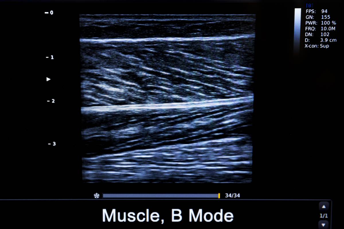
The intellectual output of an engineer-obstetrician duo, ultrasound was first used clinically as a medical imaging technology in 1956, becoming commonplace in the 1960’s, and has since established itself as a cornerstone medical imaging procedure. Key advantages over other imaging modalities include its low cost of manufacturing and use, noninvasive nature, easy visualization of hemodynamics and tissue properties, compatibility with real-time dynamic assessment, and ease of use in the form of portable ultrasound devices 1. Recently, a number of advances in musculoskeletal ultrasound imaging, in particular, have galvanized its use across multiple clinical applications 2.

First, Doppler ultrasound has been critical for the diagnosis and monitoring of inflammatory arthropathies and the assessment of neoplasms, tendinopathies, and neuropathies. For example, Doppler ultrasound can differentiate between active synovitis and chronic fibrotic synovium, as the former appears as hyperemia 3, and, in oncologic imaging, can clearly distinguish benign from malignant tumors 4.

However, traditional Doppler ultrasound remains limited by its inability to clearly visualize microvascular flow, since its algorithm filters lower velocity elements to reduce clutter. Clearly assessing microvascular flow is key, though, to diagnosing early inflammatory processes and neoplastic angiogenesis 4,5. To this end, multiple advanced ultrasound methods have been developed. First, contrast-enhanced ultrasound, combining microbubble-based intravenous contrast agents with traditional ultrasound, can, unlike previous Doppler ultrasound methods, display blood perfusion and identify capillary-level neovascularization 6. Second, another distinct Doppler technique, incorporating a unique motion suppression algorithm that isolates and eliminates clutter, can also enable the visualization of low flow microvasculature 7.

Most recently, ultra-high frequency transducers, reaching frequencies of 20-70 MHz, far exceeding the traditional 12-18 MHz range, have also been used to visualize increasingly granular anatomical detail. As such, individual fascicles may now be distinguished using the newest high-frequency ultrasound technology 8. Clinically, this first means that ultrasound-guided neurosurgical procedures can spare nerve fascicles, presenting clear advantages over conventional ultrasound or magnetic resonance imaging 9. Second, various musculoskeletal features can be accurately measured. Tendons are easily seen, thereby enabling, for example, the identification of pulleys during ultrasound-guided treatments, resulting in safer surgeries 10. Capsular ligaments, retinacula, and fasciae, all involving millimeter-diameter mesenchymal structures, are now also distinctly visible 2. Finally, this new level of precision means that ultrasounds can be used for the early detection of neoplastic masses several millimeters in size. Importantly, however, as the frequency of the ultrasound beams increase, penetration decreases. Thus, these ultrahigh frequency transducers are best suited for assessing for superficial structures.

Finally, elastography can now be used to investigate and possibly heal musculoskeletal system function. To this end, assessing stiffness of nerves and tissue around nerves in fibro-osseous tunnels can be done either by traditional strain or shear-wave elastography techniques 11.

Ultrasound is less physically and economically demanding on patients and has been greatly improving the quality of medical care. Given the rapidity of advances in ultrasound technologies, there is a clear need to standardize the execution and reporting of different associated techniques. Nonetheless, ultrasound continues to represent an indispensable diagnostic imaging modality in clinical settings, with likely many exciting additional future prospects.  

References

1.        Matsuzaki M. The latest technology of musculoskeletal ultrasonography: iterative revolution. J Med Ultrason. 2017. doi:10.1007/s10396-017-0799-0

2.        van Holsbeeck M, Soliman S, van Kerkhove F, Craig J. Advanced musculoskeletal ultrasound techniques: What are the applications? Am J Roentgenol. 2021. doi:10.2214/AJR.20.22840

3.        Aggarwal R, Aggarwal V. High-Resolution musculoskeletal ultrasound in India: The present perspective and the future. Indian J Rheumatol. 2018. doi:10.4103/0973-3698.238196

4.        Jiang Z Zhen, Huang Y Hua, Shen H Liang, Liu X Tian. Clinical Applications of Superb Microvascular Imaging in the Liver, Breast, Thyroid, Skeletal Muscle, and Carotid Plaques. J Ultrasound Med. 2019. doi:10.1002/jum.15008

5.        Arslan S, Karahan AY, Oncu F, Bakdik S, Durmaz MS, Tolu I. Diagnostic performance of superb microvascular imagingand other sonographic modalities in the assessment of lateral epicondylosis. J Ultrasound Med. 2018. doi:10.1002/jum.14369

6.        Qin S, Caskey CF, Ferrara KW. Ultrasound contrast microbubbles in imaging and therapy: Physical principles and engineering. Phys Med Biol. 2009. doi:10.1088/0031-9155/54/6/R01

7.        Yokota K, Tsuzuki Wada T, Akiyama Y, Mimura T. Detection of synovial inflammation in rheumatic diseases using superb microvascular imaging: Comparison with conventional power Doppler imaging. Mod Rheumatol. 2018. doi:10.1080/14397595.2017.1337288

8.        Cartwright MS, Baute V, Caress JB, Walker FO. Ultrahigh-frequency ultrasound of fascicles in the median nerve at the wrist. Muscle and Nerve. 2017. doi:10.1002/mus.25617

9.        Forte AJ, Boczar D, Oliver JD, Sisti A, Clendenen SR. Ultra-high-frequency Ultrasound to Assess Nerve Fascicles in Median Nerve Traumatic Neuroma. Cureus. 2019. doi:10.7759/cureus.4871

10.      Yang TH, Lin YH, Chuang BI, et al. Identification of the Position and Thickness of the First Annular Pulley in Sonographic Images. Ultrasound Med Biol. 2016. doi:10.1016/j.ultrasmedbio.2015.12.007

11.      Ooi CC, Malliaras P, Schneider ME, Connell DA. “Soft, hard, or just right?” Applications and limitations of axial-strain sonoelastography and shear-wave elastography in the assessment of tendon injuries. Skeletal Radiol. 2014. doi:10.1007/s00256-013-1695-3

The Public Health Benefits of Harm Reduction Interventions

Thousands of people suffer from substance abuse and addiction disorders all over the country and world. The typical policy response to this epidemic has been aimed at eradicating this behavior through policing and criminal justice action, and yet, no meaningful progress has been made1. Many advocates and researchers have raised the question: is this all-or-nothing approach the right way to decrease drug use and its associated problems? Or is the middle ground, “harm reduction” approach more effective?

The philosophy of harm reduction has long since been considered controversial, because it values policy favoring pragmatism over total eradication1. For example, harm reduction proponents operate under the assumption that IV drug abuse will endure, and that the focus should instead be on minimizing transmission of Hepatitis C and HIV. Another example is focusing on reducing potential opioid overdose by opening methadone clinics and having community access to Narcan, rather than eliminating harmful opioid use2. The controversy surrounding harm reduction is also likely due in part to unclear definitions and messaging, however, public health experts believe that community-driven, evidence-based harm reduction efforts do have a net positive effect on the overall health of vulnerable populations1.

The first large, real harm reduction program was established in the 1980s, in Liverpool, and was targeted at reducing the prevalence of HIV in people using IV drugs2. Program workers implemented the Mersey Model of Harm Reduction, which had three main principles: making contact with the population at risk (particularly those not already accessing healthcare), maintaining contact with those people, and making changes in their behavior2. They offered a variety of services including needle exchanges, opioid substitution therapy, STD and HIV testing programs and outreach programs to reach as many people as possible in need of help2. In the first two years, over 1000 people utilized these services and were responsible for a third of the methadone prescribed in England in that time period2. Needle exchange programs and treatment units like the one in Liverpool were set up all over the region, and it was estimated that in the next five years, about 10,000 people were able to benefit from this model2. Studies showed that the sharing of needles was significantly reduced in that area, and crime in Merseyside also went down during this time, particularly burglary and theft from vehicles, a reduction attributed to the methadone clinics set up by this program2.

Harm reduction goes beyond IV drugs; an even more common problem is tobacco use, which is associated with many health risks3. Smokeless tobacco is the leading model of THR (tobacco harm reduction) today and has been endorsed by the American Council on Science and Health as being a 98% safer alternative to smoking3. Smokeless tobacco is not associated with an elevated risk of heart attack, hypertension, and GI symptoms when compared to smoking3. Smokeless tobacco has even been proven to be safer in pregnant women, and while all forms of nicotine should be avoided in pregnancy, smokeless tobacco offers less teratogenic risk than smoking cigarettes3. While more research needs to be done to solidify smokeless tobacco as a legitimate strategy for people who smoke, these preliminary results are promising3.

Harm reduction can be a difficult pill to swallow; should we as a society settle for “less bad” instead of “good?” However, if implemented in an evidence-based fashion, these programs and models have been shown to improve overall health and outcomes, and should be considered when making health policy decisions1.

References 

  1. Ball AL. HIV, injecting drug use and harm reduction: a public health response. Addiction, 2007; 12(5): 684-690. https://doi.org/10.1111/j.1360-0443.2007.01761.x 
  1. Ashton JR, Seymour H. Public Health and the origins of the Mersey Model of Harm Reduction. International Journal of Drug Policy, 2010; 21(2): 94-96. https://doi.org/10.1016/j.drugpo.2010.01.004 
  1. Rodu B. The scientific foundation for tobacco harm reduction, 2006-2011. Harm Reduction Journal, 2011; 8. https://doi.org/10.1186/1477-7517-8-19 

The Development of Mechanical Ventilation

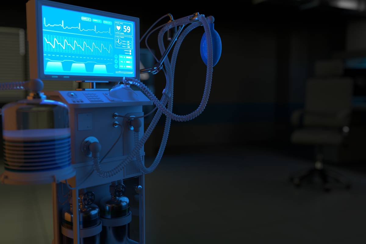
In the early days of the Covid-19 pandemic, ventilator shortages and their importance in the treatment of the disease brought renewed attention to mechanical ventilation. Ventilatory assistance has been used to help patients with airway injuries, lung damage and other conditions for centuries. However, the history of modern mechanical ventilators dates back to the mid-19th century.  

The earliest mention of mechanical ventilation comes in the writings of Vesalius, a 16th century academic. His writings on the human anatomy detailed a method of positive pressure ventilation and described a lifesaving procedure that today would be called a tracheotomy [1]. However, most of his research was hypothetical — it would take several centuries until a machine capable of mechanical ventilation was invented.  

The first mechanical ventilators were produced in the 1800s. The earliest mechanical ventilators worked by enclosing the patient in a box that contained negative pressure, which was then used to replace the patient’s respiration. In 1864, Alfred Jones created a prototype device wherein the patient’s seated body was enclosed within a sealed box. A plunger was used to create negative pressure, which would cause inhalation. When that plunger was released, the pressure would stabilize, causing exhalation [2].  

A few years later, in 1876, the first iron lung was built, but it was not widely used at first [1]. However, the poliomyelitis outbreak in the early 20th century led to a widespread need for ventilators. The virus that causes the disease can attack the respiratory system, causing respiratory failure [3]. In 1929, an electrically-powered ventilator was built and sold by Drinker and Shaw. This became the standard for treating polio in the United States [4]. Later versions allowed nurses to access the patient. 

The first positive pressure ventilators were developed in the 1950s, spurred by a new, larger outbreak of polio. At first, these ventilators were non-invasive but, due to their efficacy, invasive positive pressure ventilators were soon the norm. The early generations of ICU ventilators also offered rudimentary monitoring capabilities, which allowed physicians to monitor the patient’s tidal volume and respiratory rate [5]. The third generation of ventilators would include more robust gas delivery systems, as well as additional alarm and monitoring functions.  

The current generation of ventilators is the result of more than five decades of research into positive pressure ventilators, as well as centuries of observations regarding mechanical ventilation. These ventilators offer advanced monitoring capabilities and a host of automated features. Likewise, they also offer newer modes of ventilation, including pressure-control ventilation, synchronized intermittent mandatory ventilation, and pressure-support ventilation (PSV) [6]. However, many of the hazards of invasive ventilators that marred the early years of the technology still exist. 

Even as positive pressure ventilators continue to dominate, the pandemic has led some researchers to question whether there is still a role for negative pressure ventilators. Unlike positive pressure ventilators, negative pressure ventilators tend to cost less and have a lower risk of complications. More recent proposals are also significantly smaller in size than the iron lungs of 20th century. Regardless of the form of ventilation, future mechanical ventilators will undoubtably remain one of the most essential tools in anesthesiology, surgery, and intensive care. 

References 

[1] Slutsky, Arthur S. “History of Mechanical Ventilation. From Vesalius to Ventilator-Induced Lung Injury.” American Journal of Respiratory and Critical Care Medicine, vol. 191, no. 10, 2015, pp. 1106–1115., doi:10.1164/rccm.201503-0421pp.  

[2] The Evolution of “Iron Lungs”: 1928-1978. J.H. Emerson Co., 1978.  

[3] Wunsch, Hannah. “The Outbreak That Invented Intensive Care.” Nature, 2020, doi:10.1038/d41586-020-01019-y.  

[4] Maxwell, James H. “The Iron Lung: Halfway Technology or Necessary Step?” The Milbank Quarterly, vol. 64, no. 1, 1986, p. 3., doi:10.2307/3350003

[5] Kacmarek, R. M. “The Mechanical Ventilator: Past, Present, and Future.” Respiratory Care, vol. 56, no. 8, 2011, pp. 1170–1180., doi:10.4187/respcare.01420.  

[6] Jain, Rajnish K., and Srinivasan Swaminathan. “Anaesthesia Ventilators.” Indian Journal of Anaesthesia, vol. 57, no. 5, 2013, p. 525., doi:10.4103/0019-5049.120150.  

[7] Abughanam, Nada, et al. “Investigating the Effect of Materials and Structures for Negative Pressure Ventilators Suitable for Pandemic Situation.” Emergent Materials, vol. 4, no. 1, 2021, pp. 313–327., doi:10.1007/s42247-021-00181-x.  

Interscalene Block: Uses and Consideration

In 1970, the interscalene or brachial plexus block was devised as a technique for administering regional anesthesia of the shoulder. This technique has evolved over time, accessing the brachial plexus first through a nerve stimulator and more recently through an ultrasound guided block1.  

Administration of local anesthesia in shoulder surgery is made difficult by the emergence of the brachial plexus between the anterior scalene and middle scalene, and its subsequent, complicated descent and innervation of the shoulder and upper limb1. Because of this, general anesthesia is often preferred during shoulder surgery1. Interscalene block provides an option for regional anesthesia during surgery and, more commonly, is also used for preventing and managing postoperative pain1. The interscalene block is the most common postoperative analgesic for shoulder arthroplasty, and the PROSPECT guidelines have named interscalene block as the first line treatment for pain after rotator cuff surgery5. It can be used successfully with adjuvant therapies as well; one study showed that dexmedetomidine, an alpha-2 receptor agonist, works synergistically with an interscalene block to reduce postoperative pain after arthroscopic rotator cuff repair3. Because this combination of therapies significantly decreases postoperative pain in shoulder surgery patients, it offers an alternative to opioids in pain management3.  

However, there are some important complications of this technique that should be considered. The local anesthetic could be spread to the phrenic nerve, the sympathetic ganglia or other tissues, causing hemi diaphragmatic paresis, Horner syndrome and hoarseness2. Other acute reported complications include peripheral neuropathy caused by nerve injury and postoperative paresthesias, and while these were originally thought to be due to the block itself, researchers are investigating the possibility that they are caused by the interactions between the peripheral block and patient risk factors2. Rarely, an interscalene block can cause spontaneous pneumothorax and seizures, although it is not generally considered to be a risk factor for these complications2. Furthermore, the interscalene block has reportedly caused chronic complications in rare cases, such as carpal tunnel, prolonged paresthesia and loss of sensory function in the hand, and in one puzzling case, idiopathic neuropathy2. The patient in question had an uncomplicated interscalene block combined with general anesthesia, and their symptoms spontaneously resolved in nine months2.  

While this technique does not seem to cause long-term or unresolvable complications, the complications noted above have caused researchers to consider the use of other access techniques, such as the interscalene perineural catheter or the supraclavicular plexus block2,4. One study in the European Journal of Anesthesiology performed a systematic review and meta-analysis on the efficacy of the supraclavicular plexus block in preventing postoperative pain compared to the interscalene block4. This study found that the two analgesics resulted in comparable pain scores, but that the supraclavicular block was associated with fewer adverse events4.  

Interscalene block has provided an avenue for managing pain in patients that have undergone shoulder surgery. Clinicians should investigate the use of interscalene block on a case by case basis and evaluate risk prediction based on individual patient health profiles.  

References 

  1. Banerjee S, Acharya R, and Sriramka B. Ultrasound-Guided Inter-scalene Brachial Plexus Block with Superficial Cervical Plexus Block Compared with General Anesthesia in Patients Undergoing Clavicular Surgery: A Comparative Analysis. Anesthesia Essays and Researches2019; 13(1): 149-154. doi: 10.4103/aer.AER_185_18 
  1. Borgeat A, Ekatodramis G, Kalberer F, Benz C. Acute and Nonacute Complications Associated with Interscalene Block and Shoulder Surgery: A Prospective Study. Anesthesiology, 2001; 95: 875-880. doi: 10.1097/00000542-200110000-00015 
  1. Hwang JT, Jang JS, Lee, JJ et al. Dexmedetomidine combined with interscalene brachial plexus block has a synergistic effect on relieving postoperative pain after arthroscopic rotator cuff repair. Knee Surgery, Sports Traumatology, Arthroscopy2020; 28: 2343–2353. doi: 10.1007/s00167-019-05799-3 
  1. Schubert AK, Dinges HC, Wulf H, Wiesmann T. Interscalene versus supraclavicular plexus block for the prevention of postoperative pain after shoulder surgery. European Journal of Anaesthesiology, 2019; 36: 427-435 doi: 10.1097/EJA.0000000000000988. 
  1. Toma O, Persoons B, Pogatzki-Zahn, E, Van de Helde M, Joshi GP. PROSPECT guideline for rotator cuff repair surgery: systematic review and procedure specific postoperative pain management recommendations. Anaesthesia, 2019; 74(10): 1320-1331. doi: 10.1111/anae.14796 

Elevated D-Dimer Levels in COVID-19 Patients

D-dimer is the primary breakdown fragment of fibrin and is routinely used as a biomarker of fibrinolysis and coagulation [1]. Healthy individuals have low levels of circulating D-dimer, whereas elevated levels are frequently found in conditions associated with chronic inflammation and thrombosis [1]. Given that widespread microthrombi have been observed in multiple organ systems in patients with COVID-19, many recent studies have focused on the role of D-dimer in determining disease outcomes [1]. 

Elevated D-dimer levels have been linked to several adverse events, including thrombosis, critical illness, acute kidney injury, and all-cause mortality [1]. Patients with severe COVID-19 have a higher level of D-dimer than those with a mild infection [3]. A D-dimer level greater than 0.5 μg/ml is associated with severe infection in patients with COVID-19 [3]. Higher in-hospital mortality is associated with D-dimer levels greater than 3 μg/ml [3]. Markedly elevated D-dimer has also been linked to 28-day mortality in COVD-19 patients [3].  

The D-dimer test is commonly used in clinical practice to exclude a diagnosis of deep vein thrombosis (DVT), pulmonary embolism (PE), or disseminated intravascular coagulation (DIC) [2]. Elevated D-dimer levels indicate increased risk of abnormal blood clotting and are found in almost all patients with severe DVT [2,3]. A rise in D-dimer levels has also been associated with a higher mortality rate in community-acquired pneumonia [3]. The amount of D-dimer in the body can be measured using various commercial kits based on a monoclonal antibody [2]. The sensitivity of these D-dimer test kits ranges between 93% and 95% [2]. 

COVID-19 predisposes patients to thrombotic disease, both in venous and arterial circulation, due to excessive inflammation, platelet activation, endothelial dysfunction, and stasis [4]. Patients with COVID-19 infection are up to 25% more likely to experience thrombosis [2]. Elevated D-dimer levels are associated with the disease progression of COVID-19 and increased incidence of coagulopathy [3]. The levels of D-dimer in patients with COVID-19 admitted to the ICU has been reported to be significantly increased [3]. Patients with COVID-19 infections in the ICU are often preemptively treated with therapeutic anticoagulation in order to prevent future thrombotic events [5]. A 2020 study examined the incidence of thrombotic complications in 184 COVID-19 patients admitted to the ICU [5]. The results showed a 31% incidence of thrombotic complications, with PE ranking as the most frequent thrombotic complication [5]. 

There is also evidence to suggest that D-dimer may not only be a biomarker of hypercoagulability but also may participate in pathogenesis [1]. Fibrin degradation products have a direct procoagulant effect by inducing acute pulmonary dysfunction [1]. They also increase platelet aggregation, prostaglandin synthesis, complement activation, and initiation of chemotaxis and neutropenia [1]. Underlying conditions such as diabetes, cancer, stroke, and pregnancy may trigger an increase in D-dimer levels in COVID-19 patients [6]. 

 One of the most consistent hemostatic abnormalities associated with COVID-19 is increased D-dimer levels [4]. Although the cause of this elevation is uncertain, new data has indicated that a 3- to 4-fold rise in D-dimer levels is associated with poorer prognosis [4,6]. When treating patients with COVID-19, physicians should be aware of D-dimer level status and vigilant to signs of thrombotic complications [5]. 

References 

  1. Berger, J., Kunichoff, D., Adhikari, S. et al. (2020). Prevalence and Outcomes of D-Dimer Elevation in Hospitalized Patients With COVID-19. Arteriosclerosis, Thrombosis, And Vascular Biology, 40(10), 2539-2547. doi:10.1161/atvbaha.120.314872 
  1. Rostami, M., & Mansouritorghabeh, H. (2020). D-dimer level in COVID-19 infection: a systematic review. Expert Review of Hematology, 13(11), 1265-1275. doi:10.1080/17474086.2020.1831383 
  1. Yu, H., Qin, C., Chen, M. et al. (2020). D-dimer level is associated with the severity of COVID-19. Thrombosis Research, 195, 219-225. doi:10.1016/j.thromres.2020.07.047 
  1. Demelo-Rodríguez, P., Cervilla-Muñoz, E., Ordieres-Ortega, L. et al. (2020). Incidence of asymptomatic deep vein thrombosis in patients with COVID-19 pneumonia and elevated D-dimer levels. Thrombosis Research, 192, 23-26. doi:10.1016/j.thromres.2020.05.018 
  1. Klok, F., Kruip, M., van der Meer, N. et al. (2020). Incidence of thrombotic complications in critically ill ICU patients with COVID-19. Thrombosis Research, 191, 145-147. doi:10.1016/j.thromres.2020.04.013 
  1. Yao, Y., Cao, J., Wang, Q. et al. (2020). D-dimer as a biomarker for disease severity and mortality in COVID-19 patients: a case control study. Journal of Intensive Care, 8(1). doi:10.1186/s40560-020-00466-z